

Wall of Fame
Is your case great enough to be selected for our Wall of Fame?
If so, we’ll post your name and case here for the world to see!
AND…
Your case will be published in the PubMed-indexed Ophthalmic Surgery, Lasers and Imaging Retina.
You’ll be eligible to present at the twice yearly combined Retina Rocks – Retina Image Bank – European Vitreoretinal Society Image Bank E-Live.
Our top 3 images of the year get a free registration to present at the Retina World Congress annual meeting in Fort Lauderdale, Florida.
September 2026
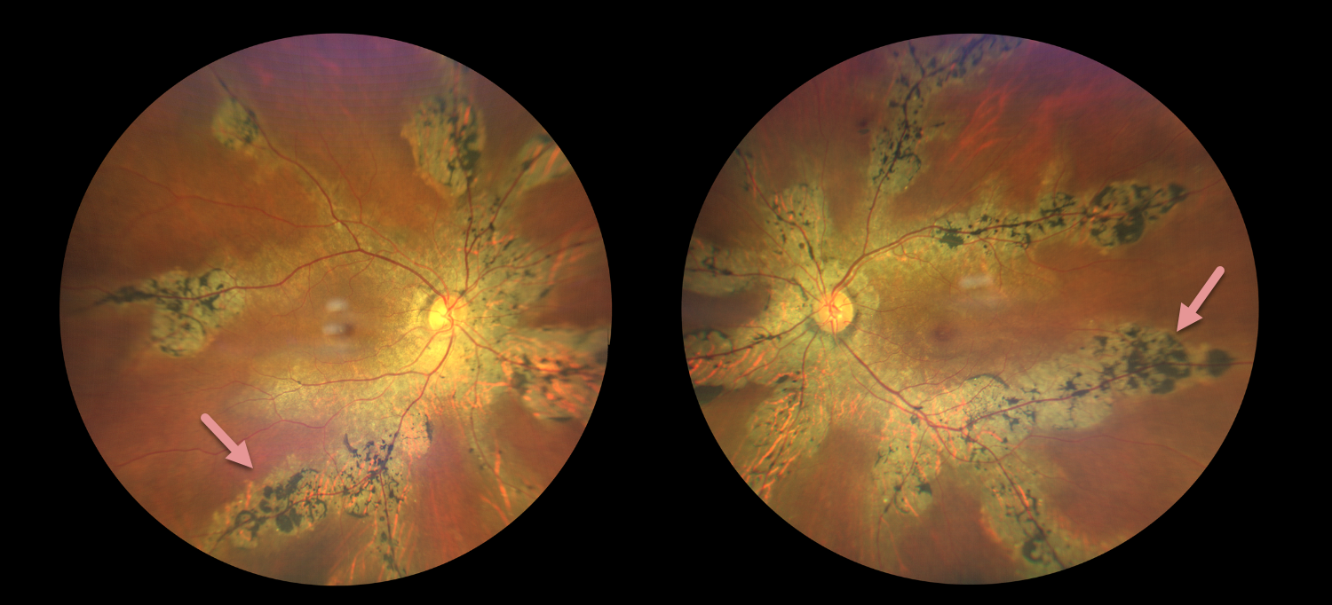
PIGMENTED PARAVENOUS CHORIORETINAL ATROPHY
Brahim KhourI, Oriana Gomez, Sebastian Polo, and Axel Ramos Nuñez Cazares
This healthy 55YO female presented with 2 years of mild bilateral vision loss. Family history was negative. Vision was 20/20 OD and 20/40 OS.
Color photography shows bilateral paravenous chorioretinal scarring with intraretinal pigment migration.
Learning Points:
Pigmented paravenous chorioretinal atrophy (PPCRA) is a rare condition characterized by paravenous pigment clumps with peripapillary and radial zones of retinal pigment epithelial (RPE) atrophy (Lee et al, AJO 2021;224:120-132). It is bilateral, often asymmetric, with various causes, including genetic and pseudo-PPCRA (inflammatory, infectious, or unknown). Patients may be asymptomatic or present with symptoms such as nyctalopia, while central visual acuity is often relatively preserved in the absence of macular involvement. The underlying pathophysiology remains incompletely understood, although primary choroidal and RPE abnormalities have been proposed to contribute to the characteristic retinal changes.
August 2026
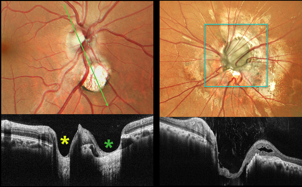
MORNING GLORY DISC AND CHOROIDAL COLOBOMA WITH PSEUDODUPLICATED OPTIC NERVE
Shraddha Raj Shrivastava and Manish Nagpal
This 26YO male presented with a history of stable poor vision and inward deviation of his left eye since childhood. Vision was 20/20 OD and counting fingers in his esotropic, microphthalmic OS.
Color photography of the right posterior pole shows a round coloboma inferonasal to the nerve, giving the appearance of a pseudo-duplicated disc. OCT scanning through the nerve and coloboma reinforces the illusion of a second nerve head. The anomalous left nerve is enlarged and excavated, with numerous vessels radiating outwards from its inferonasal aspect.
Morning glory syndrome is typically unilateral and shares some similarities with optic disc coloboma and juxtapapillary staphyloma. This abnormality is named for its striking resemblance to the Morning Glory flower. Visual prognosis is usually poor. The condition can be associated with serous macular detachment. Neuroimaging should be considered to rule out transsphenoidal encephalocele and intracranial carotid artery dysgenesis, and fortunately, our patient’s imaging was negative. For a great review on the pathogenesis and treatment of maculopathy associated with cavitary optic disc anomalies, see Jain and Johnson, AJO 2014;158:423-435.
The coexistence of morning glory disc anomaly with microphthalmos, and contralateral chorioretinal coloboma in this case, may represent a continuum of optic fissure developmental defects with asymmetric expression, leading to a spectrum of cavitary optic disc anomalies in the same individual.
Pseudoduplication of the optic nerve occurs when a round choroidal coloboma or chorioretinal scar about the size of the optic nerve is associated with overlying radiating blood vessels (Bloom et al, Retinal Cases 2022;16:174-176). In our experience, this fake-out is most common from toxoplasmosis scars located near the optic nerve.
July 2026
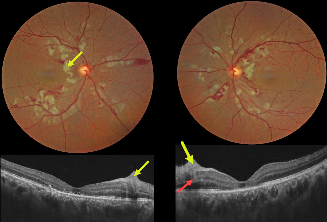
PURTSCHER RETINOPATHY
Ayushi Gupta and Vishal Agrawal
This 55YO male presented with sudden bilateral vision loss that developed immediately following a motor vehicle accident, which included blunt thoracoabdominal trauma and transient loss of consciousness. Vision was 20/200 OU.
Color photography shows bilateral inner and mid-retinal patches of white ischemic retina. The nerve fiber layer lesions appear as inner retinal hyperreflective thickening on OCT. Mid-retinal hyperreflectivity involving the outer plexiform/inner nuclear/outer plexiform layers is noted nasally OS. The outer photoreceptor bands are variably disorganized OU, with a small amount of subfoveal subretinal fluid OD and a foveal cyst OS.
Learning Points:
Purtscher retinopathy was first described by Otmar Purtscher in 1910. Although originally described in a man who fell from a tree with cranial trauma, these white patches of retinal ischemia and hemorrhages are found in numerous other etiologies, including pancreatitis, chest trauma, and collagen vascular diseases. The findings are called Purtscher retinopathy when due to trauma, and Purtscher-like retinopathy when seen from other causes.
The underlying cause of Purtscher is unknown, but it is thought to represent leukoembolization by complement activation. The areas of ischemia include more superficial nerve fiber layer infarcts (cotton wool spots) and deeper ischemia involving the middle retinal layers. This recently described retinal ischemic phenotype, paracentral middle maculopathy (PAMM), occurs as an idiopathic entity or in other ocular disorders, including Purtscher retinopathy, diabetic retinopathy, retinal artery and retinal vein occlusions, and acute macular neuroretinopathy (see Retina 2015;35:1921-1930).

June 2026
HEMORRHAGIC OCCLUSIVE RETINAL VASCULITIS (HORV)
Ayushi Gupta and Vishal Agrawal
This 35YO male underwent uneventful cataract surgery in his left eye at an outside facility about two weeks earlier. On postoperative day 4-5, he developed mild blurring and floaters, raising suspicion for early postoperative endophthalmitis. He underwent vitreous tap with intravitreal vancomycin and ceftazidime. Although he noticed initial mild symptomatic improvement, over the next 5-7 days, he experienced progressive and profound vision loss. Vision in our office was light perception. The anterior segment was quiet.
Fundus photography shows a circular band of midperipheral fibrosed retinal neovascularization encircling the posterior pole. The pericentral macula shows inner retinal white ischemia, and there are scattered retinal hemorrhages. Fluorescein angiography shows dramatic capillary loss encircling the midperiphery and temporal macula. The foveal avascular zone is irregularly enlarged. Panretinal photocoagulation (PRP) was advised, but she was lost to follow-up immediately.
Learning Points:
Hemorrhagic occlusive retinal vasculitis (HORV) is a rare but devastating complication strongly associated with intraocular vancomycin exposure, typically presenting 1-21 days after cataract surgery or intravitreal injection with delayed-onset painless vision loss (Witkin et al, Ophthalmology 2017;14:583-595). It is characterized by sectoral ischemia with retinal hemorrhages, mild anterior chamber and vitreous inflammation, often with a deceptively unremarkable postoperative day 1 examination. The findings are likely due to a delayed hypersensitivity reaction rather than direct toxicity (Todorich et al, AJO 2018;188:131-140).
The visual prognosis is poor, with approximately 61% of eyes achieving <20/200 vision and 22% progressing to no light perception. Neovascular glaucoma develops in approximately 56% of cases due to extensive retinal ischemia. Management includes early systemic and/or intravitreal corticosteroids, and subsequent PRP and anti-VEGF therapy.
May 2026
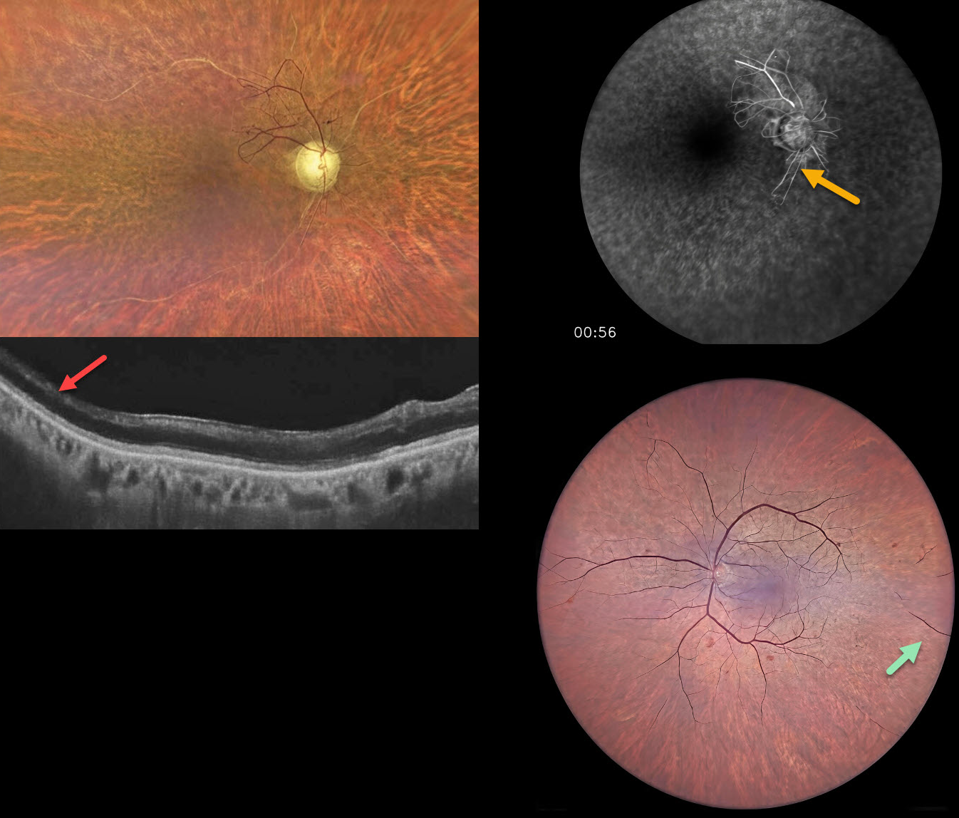
RADIATION RETINOPATHY
The European VitreoRetinal Society (EVRS) and Abhishek Upadhyaya
This 65YO male presented for evaluation for cataract surgery. He underwent external beam radiation for throat cancer, predominantly on the right side, 20 years earlier. Vision was hand motion OD and 20/200 OS.
Color photography OD shows severe panretinal ischemia with the major vessels showing absent blood columns except for some abnormal preserved vessels with anastomoses just superotemporal to the nerve. OCT shows temporal inner retinal atrophy. The left posterior pole appears more normal with severe more peripheral ischemia. Fluorescein angiography confirms the profound ischemia noted funduscopically.
Learning Points:
Radiation retinopathy usually occurs 3 months to 3 years following external beam radiation or plaque radiotherapy. The ocular findings closely resemble those seen with diabetic retinopathy. Radiation optic neuropathy can also develop (Carey et al, Br J Ophthalmol 2023;107:743-749) .
Panretinal photocoagulation was recommended for the right eye.
April 2026
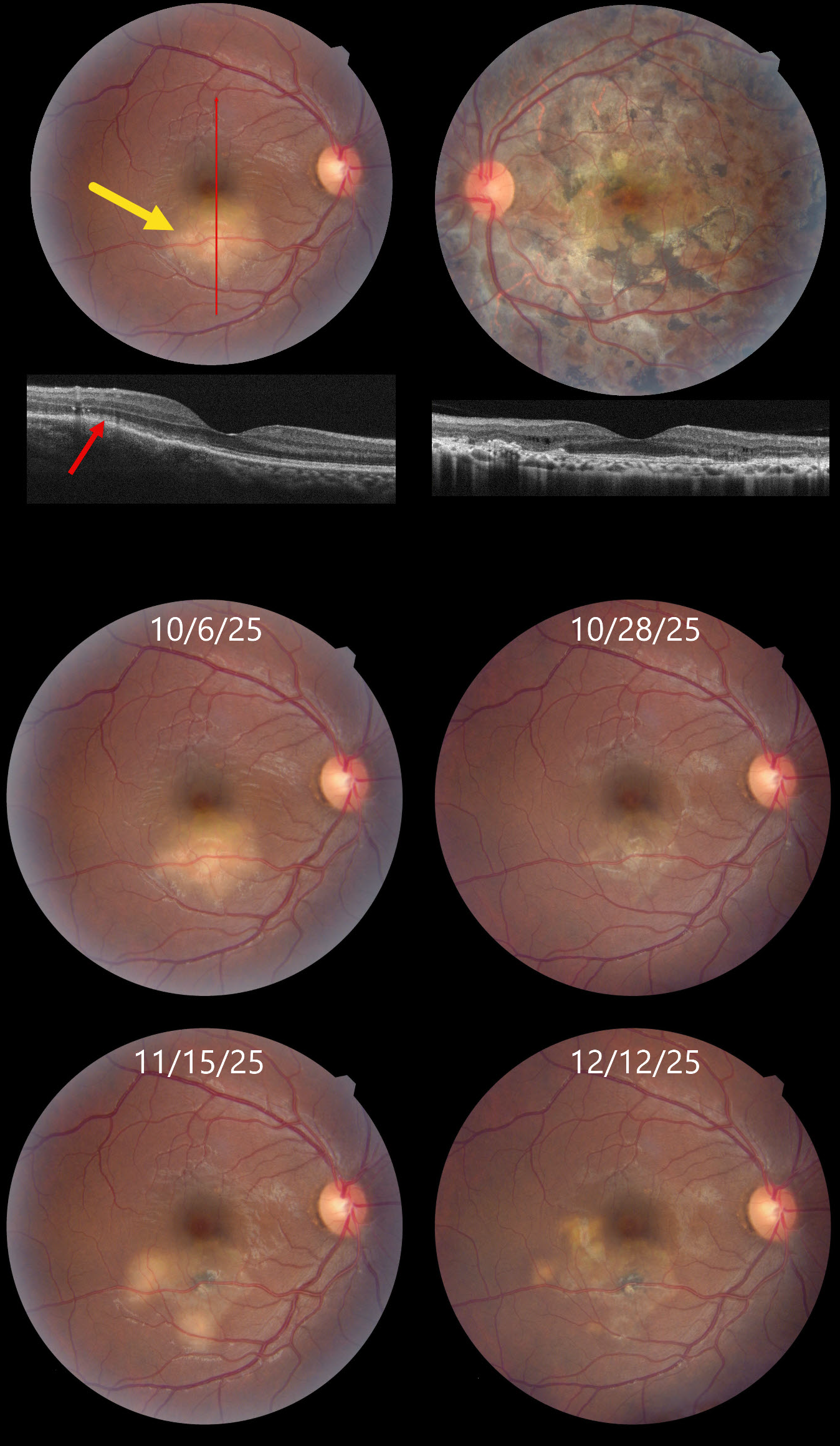
TUBERCULAR SERPIGINOUS-LIKE CHOROIDITIS
Akansha Sharma
This 32YO male presented on 10/6/25 with a 6-day history of decreased vision in his right eye. About 2 years earlier, he was diagnosed elsewhere with tubercular serpiginous-like choroiditis in his left eye. Extensive workup at the time, including a chest X-ray, was negative except for a positive QuantiFERON TB Gold result. He was lost to follow-up without treatment for his latent tuberculosis (TB). Vision in our office was 20/30 OU.
Color photography shows a deep, creamy-white retinal inflammatory lesion in the inferior macula. OCT shows outer retinal hyperreflectivity with loss of the outer retinal band details. Extensive scarring is noted throughout the left posterior pole, with a small island of preserved foveal tissue.
Tapering systemic corticosteroid therapy was begun, followed by anti-tuberculosis treatment. Over the ensuing 2 months, he experienced waxing and waning inflammation necessitating intravitreal Ozurdex, oral steroids, and adalimumab while vision remained stable at 20/30. We continue to follow him closely.
Learning Points:
Although TB most commonly presents with pulmonary involvement, extrapulmonary sites can include the gastrointestinal, skin, cardiovascular, genitourinary, and central nervous systems, as well as the eyes. Ocular involvement, like syphilis, can mimic virtually any type of uveitis, including anterior, intermediate, posterior, and panuveitis, retinitis, and retinal vasculitis, neuroretinitis, optic neuropathy, choroidal granuloma, choroiditis, and scleritis.
Tubercular serpiginous-like choroiditis mimics serpiginous but affects younger patients with more multifocal and peripheral recurrences and progression if untreated (Agarwal et al, AJO 2020;220:160-169). Although the exact mechanism is not yet clear, a direct or indirect infectious trigger by the Mycobacterium tuberculosis is thought to cause choroiditis. Its origin is probably immunogenic since it seems to respond to corticosteroids and other immunosuppressants. Our patient was a bit unusual in that he required systemic steroids, ATT, and adalimumab to finally control his disease.
March 2026
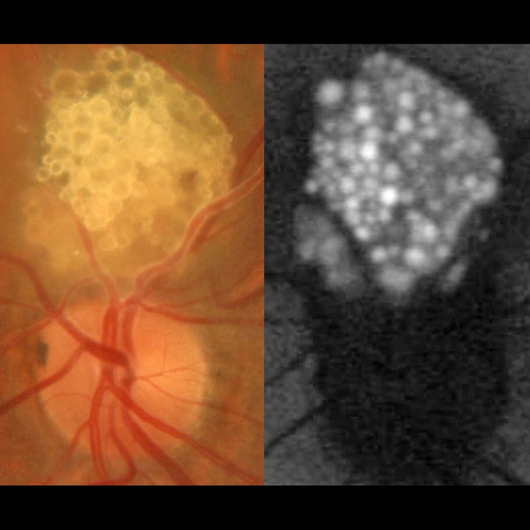
RETINAL ASTROCYTIC HAMARTOMA
European VitreoRetinal Society (EVRS) Dhaivat Shah, Nida Khan and Choithram Netralaya
This healthy 43 YO male presented with an asymptomatic lesion in his left eye. Family history was negative. Vision was 20/20 in his normal OD and 20/20 OS.
Color photography shows a glistening yellow-white inner retinal lesion just superior to the optic nerve composed of innumerable semi-transparent spherules which hyper-autofluoresce.
Learning Points:
A retinal astrocytic hamartoma is a benign tumor composed of spindle-shaped fibrous astrocytes located in the nerve fiber layer, most commonly associated with tuberous sclerosis. Tuberous sclerosis includes the classic triad of seizures, mental deficiency and fibroangiomas, but patients can have lesions throughout the body including the eyes, skin (ash-leaf spots), bone, nails, teeth, brain, heart, lungs and kidneys. Early in life the inner retinal lesions have a fleshy white appearance and are often multifocal and bilateral. With time, as in this patient, they show a more classic “mulberry appearance” with significant calcification. Rarely, the vascular component can cause intraretinal exudation requiring treatment.
Clinically retinocytomas can resemble astrocytic hamartomas. However, retinocytomas often have underlying RPE changes and chorioretinal scarring (lacking in our patient), which are absent in astrocytic hamartomas.
Since the patient was self-paying, neurologic imaging was not performed and observation was recommended. A neurology reference was given.
February 2026
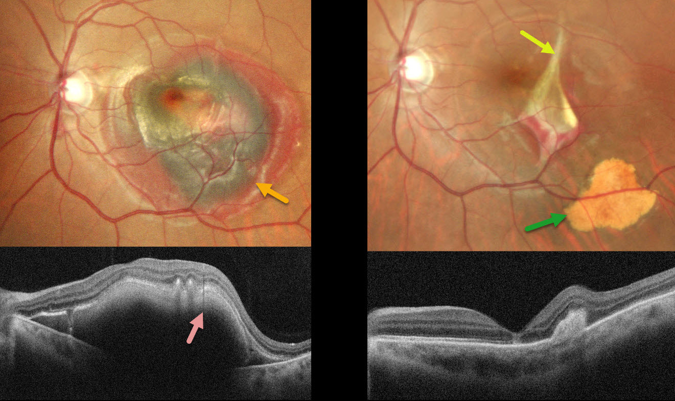
CHOROIDAL RUPTURE
Malvika Singh and Manish Nagpal
This 38YO male was hit by a stone in his left eye two days earlier. Vision was 20/20 in his normal OD and counting fingers OS.
Pseudocolor SLO imaging shows central macular greenish-brown subretinal blood surrounded by a rim of thinner red blood. OCT shows the hyperreflective blood with posterior shadowing. An in-office intravitreal C3F8 gas injection was performed, followed by face-down positioning. Six weeks later, the blood is virtually resolved except for some residual distal inferotemporal yellow devitalized subretinal blood. The causative choroidal rupture can be seen just temporal to the foveal center.
Learning Points:
An acute submacular hemorrhage following blunt trauma almost always indicates an underlying choroidal rupture as the source of the blood. The final vision is usually good unless the rupture extends through the macular center. These patients need long-term follow-up due to the increased risk of macular neovascularization.
January 2026
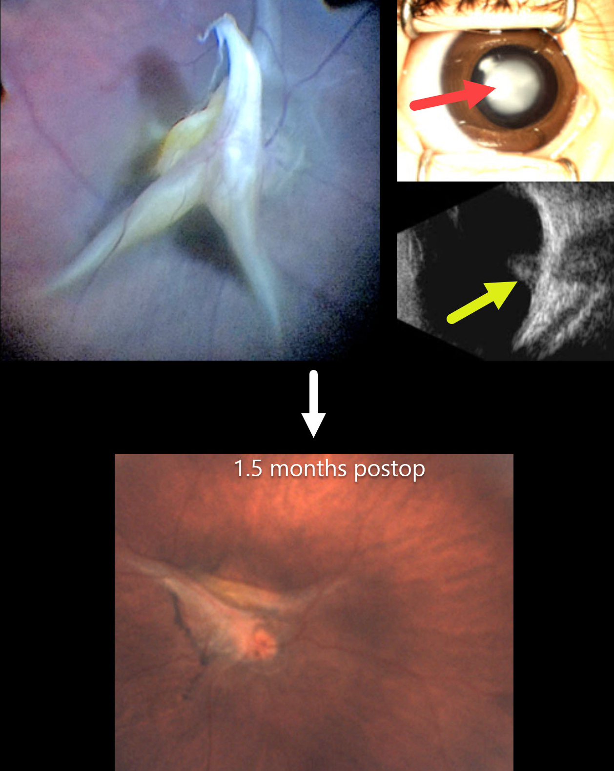
PERSISTENT FETAL VASCULATURE
European VitreoRetina Society (EVRS), Gökçen Deniz and Şengül Özdek
This healthy 4-month-old girl, born full-term, presented with leukocoria in her right eye that her parents recently noticed.
Preoperatively, there was a white central cataract with a clear peripheral zone. B-scan ultrasonography showed a highly reflective stalk that extended from the disc to the periphery with a tent-like elevation over the disc. The left eye was normal.
At surgery, the lens was aspirated with vitrectomy to release all traction. Although the stalk overlying the nerve was trimmed, persistent blood flow was noted as stalk pulsations. The retina remained attached 1.5 months postoperatively.
This case was submitted by the European VitreoRetina Society (EVRS), Gökçen Deniz, and Şengül Özdek .
Learning Points:
Persistent fetal vasculature (PFV), previously recognized as persistent hyperplastic primary vitreous (PHPV), represents a failure of regression of fetal vessels. It is a common cause of leukocoria and subsequent amblyopia. There is no identifiable genetic association.
In PFV, the arterial blood supply from the disc to the anterior segment that is supposed to regress after birth does not. This results in a remnant of the fetal hyaloid artery remaining abnormally patent. The arterial flow from this remnant can cause cataract, intraocular hemorrhages, and retinal detachment, as the abnormal vessel continues to be supplied by systemic arterial pressure.
The main surgical pearl of this case is that, when you have a clear zone around the central opacity of PFV, such eyes usually do not have peripheral retinal extensions through the ciliary body, which allows safe surgical entry through the pars plicata. One still needs to check internally that the incisions are anterior to the peripheral retina, and limbal entry is needed.
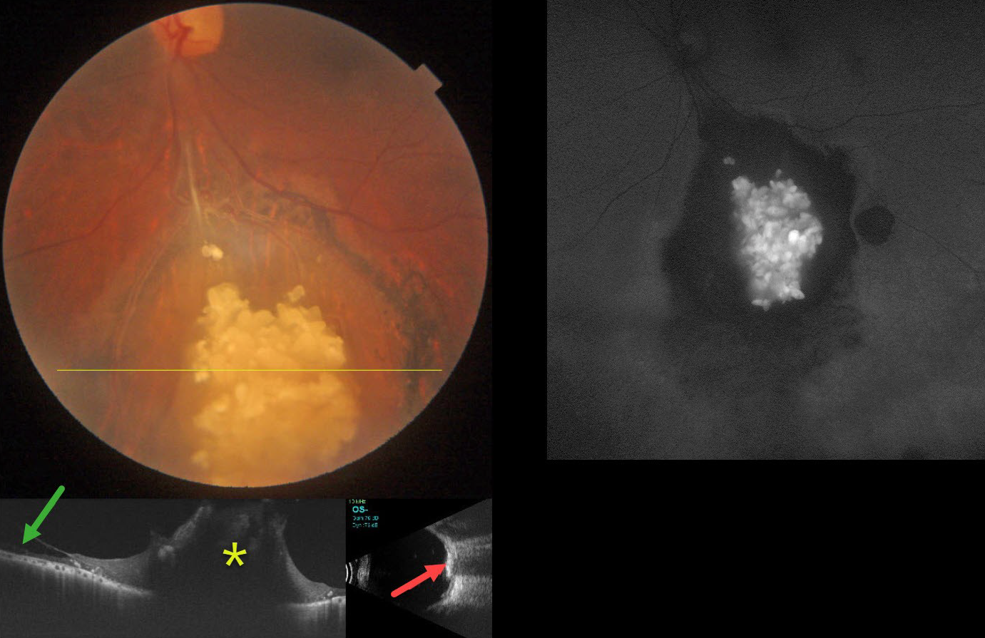
December 2025
RETINOCYTOMA
The European VitreoRetinal Society and Dibya Prabha
This healthy 43YO male presented with gradual bilateral blurring. Vision was 20/20 in his normal OD and 20/30 OS.
Color photography shows an elevated, glistening, inner retinal yellow-white mulberry-like lesion in the inferior midperiphery. Surrounding subretinal pigmentary changes are noted. OCT through this lesion shows a markedly thickened, disorganized, hyperreflective retina. Retinoschisis is noted nasally. The lesion is markedly hyperreflective on B-scan ultrasonography and hyper-autofluorescent on fundus autofluorescence.
Learning Points:
Retinocytomas are fleshy, opalescent, benign tumors that are felt to represent spontaneously regressed retinoblastoma (RB). They appear identical to previously radiated RB tumors. The risk of a germline RB1 mutation in sporadic unilateral retinoblastoma or retinocytoma is about 10-20%.
Clinically, retinocytomas can resemble astrocytic hamartomas. However, retinocytomas often have underlying RPE changes and chorioretinal scarring (as in our patient), which would not be present with astrocytic hamartomas.
The brain MRI was normal, and observation was recommended.
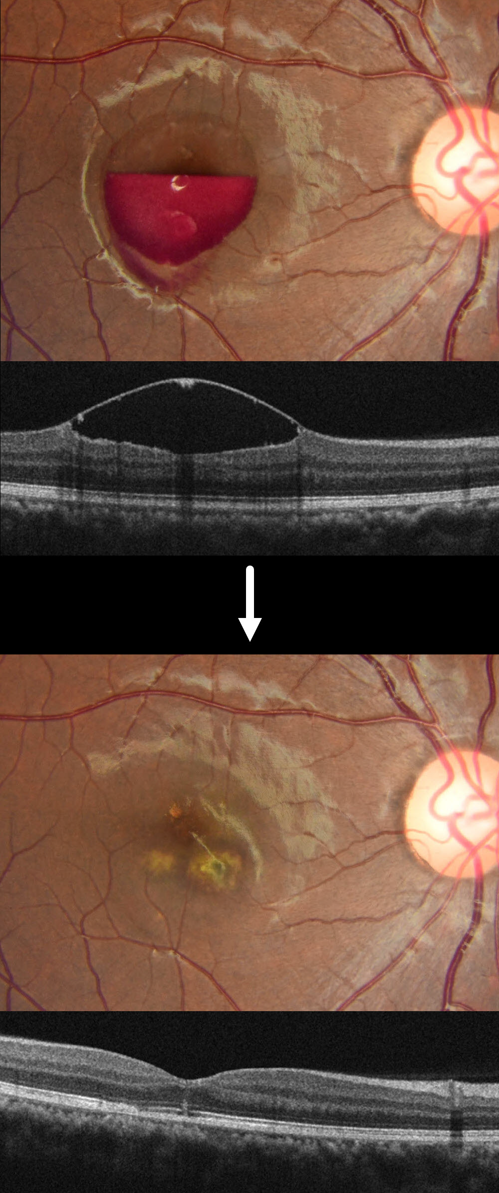
November 2025
LASER MACULOPATHY
The European VitreoRetina Society (EVRS) and Shraddha Chandorkar
This healthy 19YO female noted sudden vision loss in her right eye immediately after looking directly at the light while attending a DJ laser party. When she presented to us 2 days later, her vision was hand motion OD and 20/20 in her normal OS.
Fundus photography and OCT show a partially layered central macular sub-internal limiting membrane (ILM) hemorrhage.
When examined 1 week later, the blood was drained via a YAG laser due to no improvement in her vision or sub-ILM blood. Two months later, vision improved to 20/20 despite multiple paracentral scars.
Learning Points:
DJ laser machines typically fall into Class 3R or Class 4, depending on their power output. These lasers emit visible light in the red (635-650 nm), green (520-532 nm), and blue (445-460 nm) wavelengths, often combined for full RGB effects. While visually stunning, high-powered lasers, especially Class 4, can pose serious risks, including retinal burns, if beams enter the audience area or are viewed directly. Blue wavelengths are particularly hazardous due to their shorter wavelength and higher energy density. A strikingly similar case to our patient was reported by Perz-Montano et al (Doc Ophthalmol 2019;138:71-76).
To protect audiences, professional setups use a combination of engineering controls and regulatory compliance. This includes fail-safe scanning systems, beam termination zones, and power-limiting protocols to remain below the Maximum Permissible Exposure. In the US, the FDA’s Center for Devices and Radiological Health oversees these devices, requiring manufacturers to submit safety documentation and operators to file a Laser Light Show Variance for public performances. Proper mounting, trained personnel, and adherence to ANSI and IEC standards are essential to ensure safe and compliant laser shows.
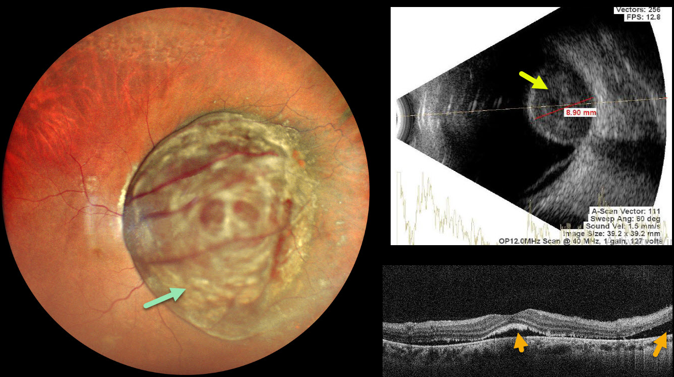
October 2025
UVEAL MELANOMA
Ankit Jain and Manish Nagpal
This 47YO female presented with 1 month of decreased vision in her right eye. Vision was 20/400 OD and 20/20 in her normal OS.
Pseudocolor SLO imaging shows a large, well-circumscribed pigmented choroidal lesion that obscures the entire posterior pole. The tumor vessels are markedly dilated and tortuous. OCT shows foveal and nasal subretinal fluid with ragged shed outer segments along the posterior retinal surface. The lesion, which measures 8.9mm in thickness, shows moderate homogenous internal reflectivity on B-scan ultrasonography. She was referred to ocular oncology for further evaluation and treatment.
Learning Points:
Uveal melanoma is a malignant tumor arising from melanocytes in the uveal tract (iris, ciliary body, or choroid). The choroid is the most common site, accounting for 90% of uveal melanomas. Uveal melanoma is the most common primary intraocular cancer in adults, whereas metastatic disease is the most common intraocular malignancy. The liver is the most common metastatic site, although metastatic disease is rare on presentation. Metastases are more common with large tumors, BAP1 mutations, and certain tumor-related genetic markers (including positive PRAME and Class 2).
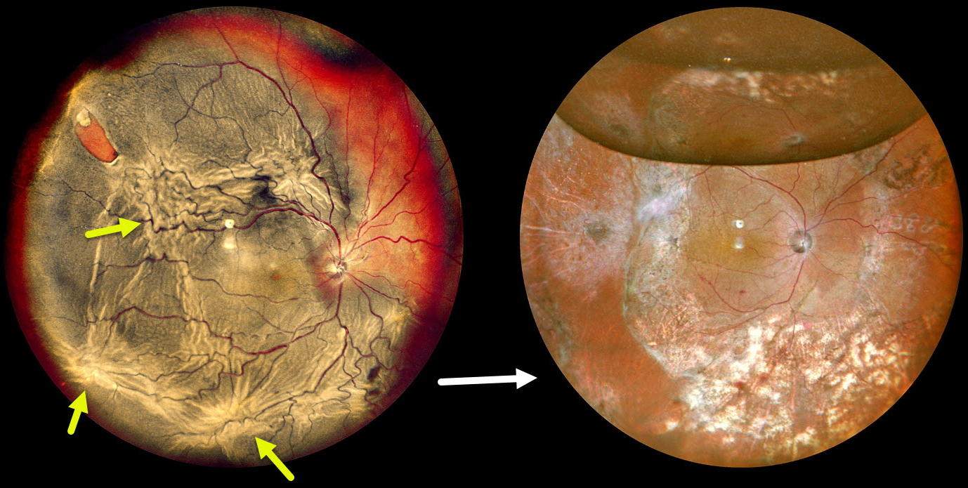
September 2025
PROLIFERATIVE VITREORETINOPATHY
The European VitreoRetina Society (EVRS) and Shishir Verghese
This 75YO male presented with decreased vision in the right eye for four months. Vision was hand motion OD and 20/25 in his normal OS.
Pseudocolor SLO imaging shows a complex total rhegmatogenous retinal detachment with a supertemporal tear and multiple proliferative vitreoretinopathy (PVR) star folds. Following three vitrectomy surgeries, including a relaxing retinectomy, the retina is attached. Vision was 20/120.
August 2025
MACULAR PUCKER WITH LAMELLAR DEFECTS
The European VitreoRetina Society (EVRS) and Shishir Verghese
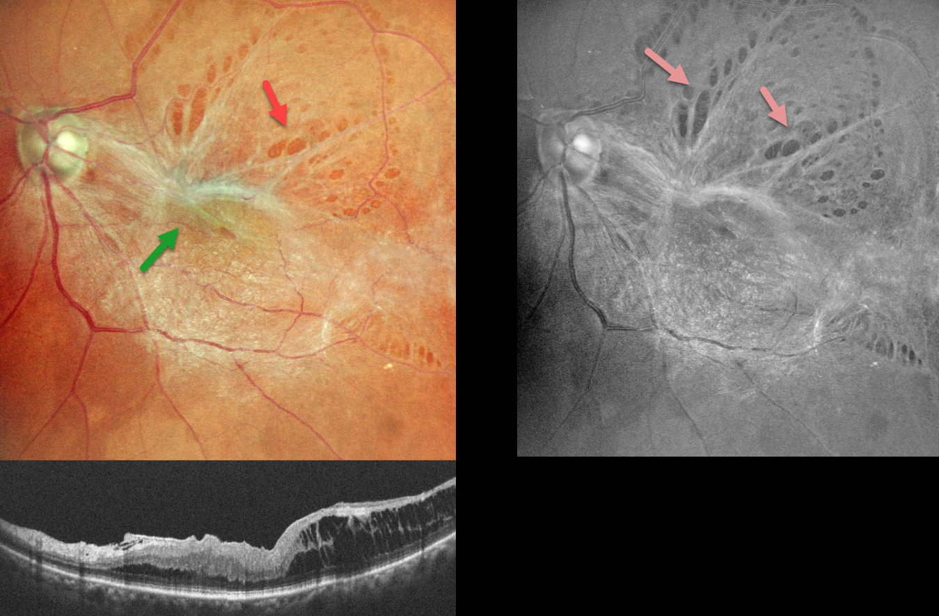
This 58YO female presented with 6 months of vision loss in her left eye. Vision was 20/60 OS.
Color SLO imaging shows a taut epiretinal membrane (ERM) with multiple paravascular lamellar defects, which are better visualized on red-free imaging. OCT scanning shows a stage 3 macular pucker with ectopic inner foveal layer (EIFL) and temporal inner layer schisis.
Learning Points:
An ERM is a thin, fibrous membrane that forms on the retinal surface, often causing central vision loss with metamorphopsia. It can be classified into 4 OCT-based stages (Gonzalez-Saldivar et al, Retina 2020;40:710-717): Stage 1 ERM with preserved foveal contour, Stage 2 Loss of foveal depression with thickened outer nuclear layer, Stage 3 EIFL, and Stage 4 EIFL with complete loss of inner retinal anatomy.
ERMs can cause paravascular lamellar defects due to shearing forces at the vessel borders, and these defects can extend towards the fovea. Our patient’s highly contractile membrane caused multiple striking lamellar defects visualized on fundus photography.
July 2025
RETINOPATHY OF PREMATURITY
Kanwaljeet Harjot Madan
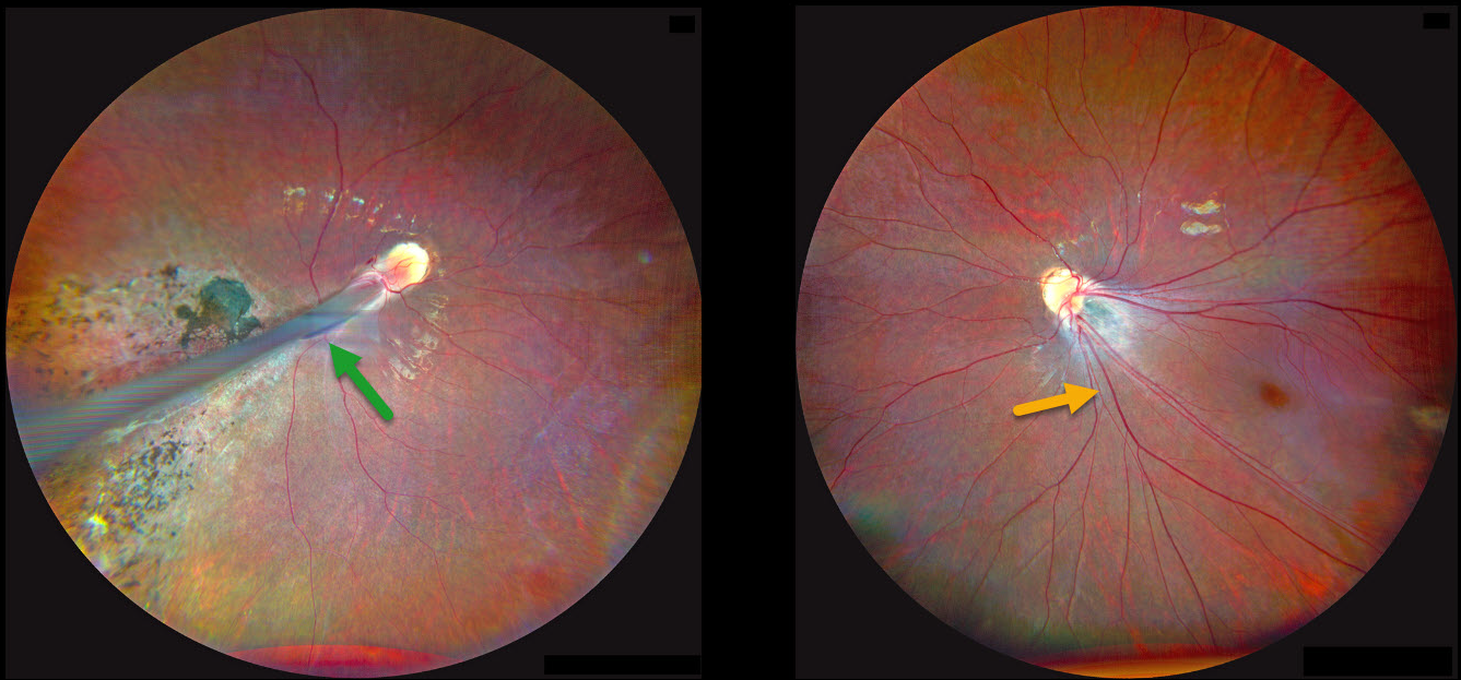
This 4YO girl’s parents noticed that her right eye was deviating outwards for several months, along with decreased vision bilaterally. Her mother gave a history of prematurity at 28 weeks of gestational age with a low birth weight of 1175 g and oxygen exposure. Vision was counting fingers OD and 20/80 OS. There was a large right exotropia.
Fundus photography OD shows a prominent retinal fold extending from the optic disc into the inferotemporal periphery. Diffuse chorioretinal scarring is noted surrounding the retinal fold. The retinal vessels OS are dragged inferotemporally with marked foveal ectopia.
Learning Points:
Retinopathy of prematurity (ROP) is a potentially blinding vasoproliferative disease occurring in premature infants. Although multifactorial, current screening guidelines are based primarily on low birth weight and gestational age (Kim et al, Surv Ophthalmology 2018;63:618-637). Screening and treatment for these eyes is extremely subspecialized, with treatment options primarily including timely scatter laser photocoagulation and intravitreal anti-VEGF injections (Yonekawa et al, Retina 2017:37:2208-2225).
Patients with ROP have an increased risk for vision-threatening complications throughout their life, including vitreous hemorrhage and retinal detachment (Hsu et al, Ophthalmology Retina 2023;7:72-80). We will therefore monitor our patient yearly.
June 2025
WELDING ARC MACULOPATHY
Rohan Jain and Manish Nagpal
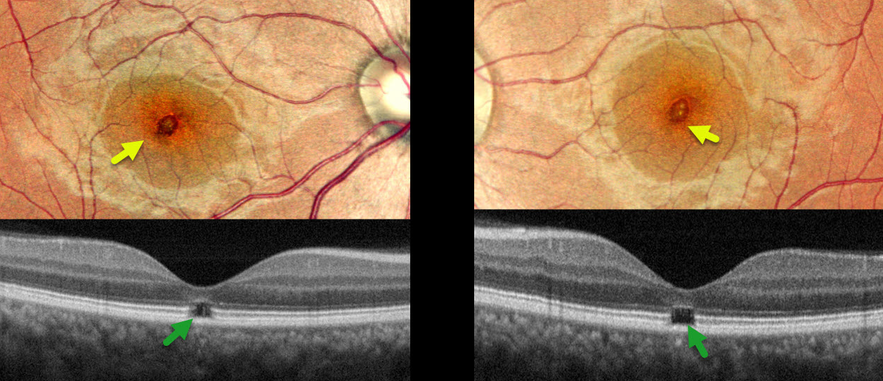
This 38YO male presented with 6-7 months of bilateral vision loss. He works as a welder and admits to rarely wearing his safety glasses. Vision was 20/25 OD and 20/20 OS.
Pseudocolor SLO imaging shows symmetrical, tiny yellow foveal scars. OCT shows outer foveal microdefects with hyporeflectivity due to loss of all bands deep to the external limiting membrane.
Learning Points:
The welding process emits optical radiation across various wavelengths and intensities, including infrared, visible, and ultraviolet (UV) light. Exposure to this UV light without proper eye protection can result in keratoconjunctivitis (the most common clinical presentation) and foveal lesions similar to those seen in solar retinopathy. Findings include a small foveal yellowish spot or scar with an outer foveal microdefect on OCT (Singh et al, Surv Ophthalmology 2023;68;655-668). Vision is usually good despite the persistent clinical and OCT findings.
Our patient was lucky to have good vision despite his poor work habits. He was advised to use protective eyewear during welding.
May 2025
POST-VIRAL FROSTED BRANCH ANGIITIS
Ayushi Gupta and Vishal Agrawal
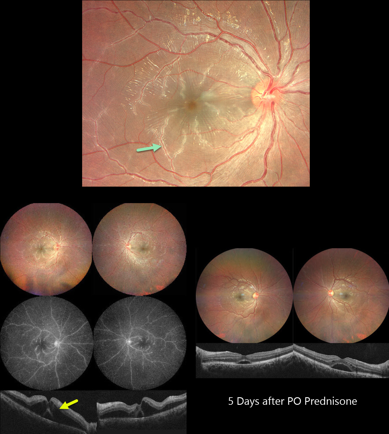
This previously healthy 21YO male developed bilateral vision loss 1 week following a viral illness. Vision was 20/100 OU.
Color photography shows a bilateral frosted-branch appearance of the retinal veins, most prominent in the maculas. Fluorescein angiography shows diffuse leakage and staining of the venous vascular tree. OCT scanning shows marked bilateral outer nuclear layer edema with foveal subretinal fluid. He was started on oral prednisone 60mg daily. Five days later vision improved to 20/40 OU. The venous changes are dramatically better with decreasing macular fluid. A slow steroid taper began.
Learning Points:
Frosted branch angiitis is a retinal vasculitis that gets its name from the involved vessels resembling branches of a tree that are “frosted” with snow (Kleiner et al, AJO 1988;106:27-34). Although these findings may be isolated, they are often considered a phenotype found in many inflammatory conditions, most commonly CMV retinitis. The inflammation is usually highly responsive to steroids, and the visual prognosis is generally good, although some may develop secondary neovascularization in the anterior and posterior segments due to widespread ischemia.
April 2025
TAMOXIFEN RETINOPATHY
Mauli Shah and Alay Banker
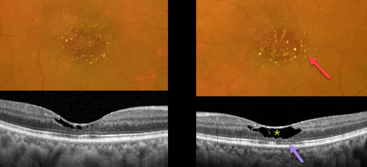
This 51YO female presented with 3 months of bilateral vision loss. She was diagnosed 5 years earlier with breast cancer, followed by a radical mastectomy. Since then, she has been treated with tamoxifen 20mg PO daily. Vision was 20/60 OD and 20/40 OS.
Color photography shows bilateral, symmetric, central macular yellowish crystalline deposits, corresponding to tiny hyperreflective inner retinal lesions on OCT. Central inner retinal cavitations, outer nuclear layer loss, and disruption of the ellipsoid zone and outer segment bands are noted, particularly in the left eye.
Learning Points:
The prevalence of tamoxifen retinopathy for patients taking tamoxifen 20mg daily may be as high as 12% (Kim et al, Ophthalmology 2020;127:555-557). Although most oncologists do not require this, it is not unreasonable to perform ophthalmic screening examinations with OCT following 2 years of therapy (Tenney et al, Surv Ophthalmology 2023;69:42-50).
Tamoxifen inhibits the glutamate-aspartate transporter, leading to excessive intracellular accumulation of glutamate in Müller cells, which are vital in maintaining retinal cell integrity and homeostasis. Tamoxifen retinopathy shows findings very similar to macular telangiectasia type 2 (MacTel 2; Lee et al., Ophthalmology Retina 2020;3:681-689), including retinal cavitations, right-angle venules, and inner retinal crystals. Both disorders likely share Muller cell injury as a common etiology. The changes in MacTel2 are usually in the temporal fovea, whereas they seem to be more diffusely distributed throughout the central macula with tamoxifen (Hess et al, Retina 2023;7:101-110).
Ocular toxicity is more common with a cumulative dose over 100 grams, although our patient still developed classic toxicity despite a cumulative dose of only 35.2 grams. The tamoxifen was discontinued after discussion with the treating oncologist. Three months later, the vision and retinal findings were unchanged (not shown).
Case of the Year
March 2025
GIANT MACULAR HOLE WITH TOTAL RETINAL DETACHMENT
Anand Temkar and Manish Nagpal
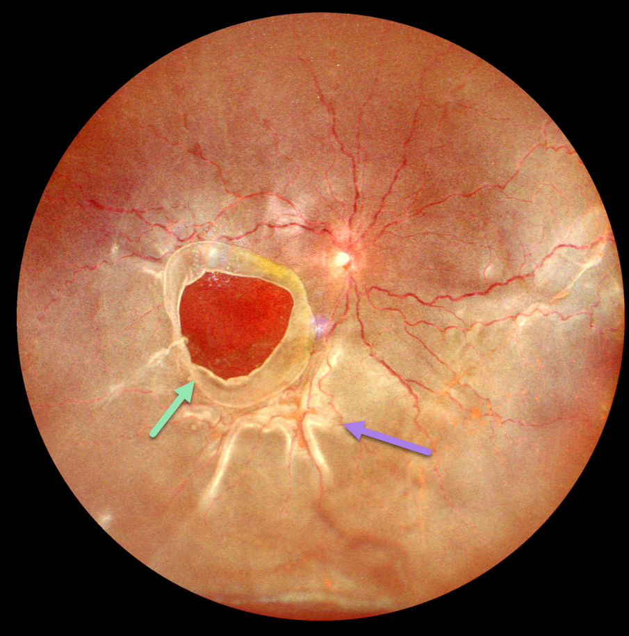
This 10YO boy presented with 1 week of vision loss in his left eye. The parents gave a vague history of trauma with a tennis ball 2 years earlier. Vision was counting fingers OD and 20/20 in his normal OS.
Ultrawidefield imaging shows a giant macular hole occupying nearly the entire macula. The hole’s edges are rolled, and proliferative vitreoretinopathy with star folds is present within the total rhegmatogenous retinal detachment (RRD).
Emergent vitrectomy surgery with silicone oil was scheduled.
Learning Points:
Blunt ocular trauma can result in numerous sequelae, including commotio retina, retinal breaks and detachment, choroidal rupture, and macular hole. A giant macular hole, also reported in Alport syndrome (Shah and Weinberg Ophthalmic Genetics 2010;31(2):93-97), is an exceedingly rare traumatic event (Hernandez-Da Mota Case Rep Ophthalmol 2011;2:283-286).
Traumatic macular holes are thought to result from a vitreous coup contrecoup injury or a jet stream of anterior chamber aqueous humor shooting through Cloquet canal into the premacular bursa.
February 2025
DEGENERATIVE RETINOSCHISIS
Vaibhav Sethi
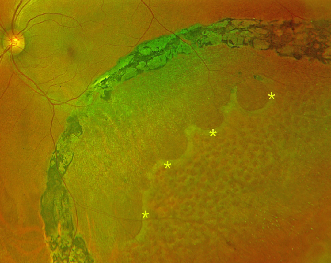
This 47YO female presented with 4 years of flashes in her left eye that began immediately following laser photocoagulation elsewhere. Vision was 20/20 bilaterally.
Optos color RG imaging shows inferotemporal degenerative retinoschisis, demarcated by heavy, confluent laser scarring along its posterior extent. A row of outer-layer holes is noted within the schisis cavity, with innumerable inner-layer holes more anteriorly.
Learning Points:
Degenerative retinoschisis is virtually never prophylactically treated (Byer 1986;93:1127-1137). Although the schisis cavities can enlarge, prophylactic treatment including thermal laser does not prevent further spread, should not be performed (Ness et al, Surv Ophthalmology 2022;67:892-907), and in our patient’s case, caused persistent symptomatic flashes. Vitrectomy surgery can successfully repair combined schisis rhegmatogenous retinal detachment (RRD) when liquid vitreous enters the subretinal space through holes in both the inner and outer layers.
December 2024
DIFFUSE UNILATERAL SUBACUTE NEURORETINITIS (DUSN)
Sharat Hegde
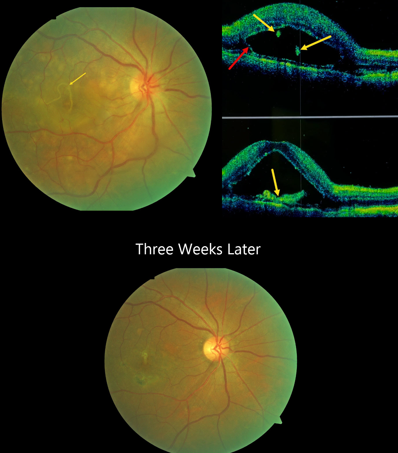
This 41YO male presented with 4 days of severe vision loss in his right eye. Vision was hand motion OD and 20/20 in his normal left eye.
Color photography shows multiple focal yellow-white deep retinal lesions with a submacular worm and overlying neurosensory detachment. OCT scanning shows a bacillary layer detachment with various cuts through the hyperreflective worm.
Direct photocoagulation of the worm was impossible due to its submacular location and continued movement. The worm subsequently died and was absorbed following a 2-week course of oral albendazole 400mg BID and steroids. Three weeks later, vision improved to 20/30. Chorioretinal scarring is noted in the region of the prior worm, and the inflammatory lesions are mostly resolved.
Learning Points:
Originally described by Gass (Ophthalmology 1978;85:521-545), diffuse unilateral subacute neuroretinitis (DUSN) is a syndrome in healthy young adults characterized by unilateral decreased vision, vitritis, and papillitis, with recurrent crops of transient gray-white outer retinal/RPE lesions. Optic atrophy, diffuse RPE changes, decreased visual field, and electroretinogram are long-term sequela in untreated cases.
DUSN is caused by at least 2 different-sized subretinal nematodes that can remain viable in the eye for up to 3 years. If identified, the extramacular worm can be treated with thermal photocoagulation, surgically removed, or treated with systemic anthelmintic therapy.
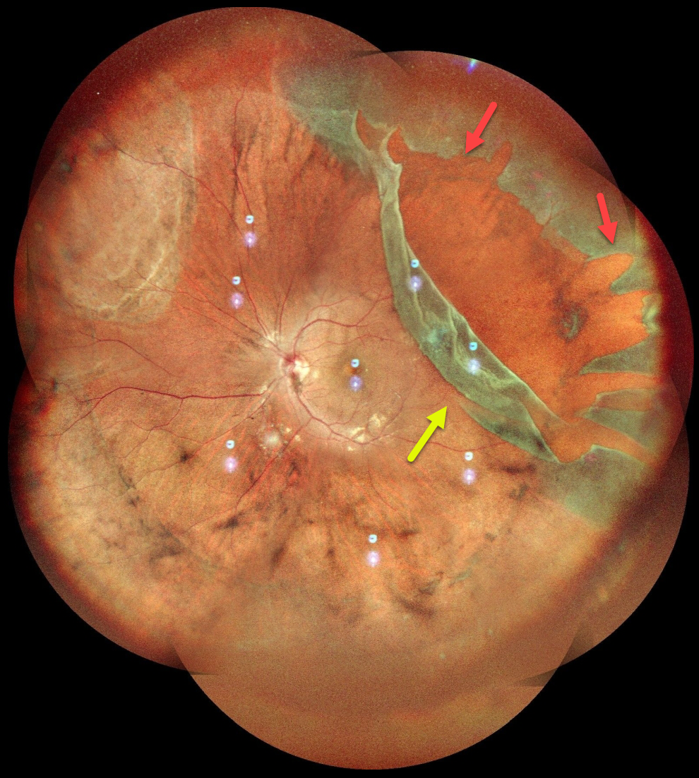
January 2025
GIANT RETINAL TEAR
Anjana Mirajkar and Manish Nagpal
This healthy 24YO male presented with several days of floaters in his left eye. Vision was 20/20 OU.
Pseudocolor SLO imaging shows a superotemporal giant retinal tear (extending over 3 clock hours) and detachment. The anterior portion of the tear has multiple jagged edges, and the posterior flap has folded over onto itself. The macula is attached. The detachment was repaired with emergent pars plana vitrectomy.
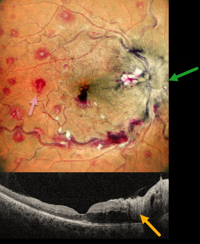
November 2024
ANEMIA
Anand Temkar and Manish Nagpal
This 35YO male presented with 1 month of vision loss in his right eye. There was no past medical history. Vision was 20/40 OD and 20/20 in his normal OS.
MultiColor imaging shows a markedly swollen optic nerve with extensive multifocal deep, white-centered retinal hemorrhages. OCT shows marked mostly outer retinal edema extending from the nerve towards the macular center.
Extensive laboratory evaluation was negative except for a microcytic anemia with a hemoglobin of 2.9 g/dl. This was felt to be caused by an iron deficiency due to poor diet. Intravitreal anti-VEGF injections were recommended, along with oral prednisone for the optic nerve swelling. Unfortunately, he was immediately lost to follow-up.
Learning Points:
Anemia-related retinal findings most commonly include hemorrhages in the retina and sub-internal limiting membrane. Severe anemia is a rare cause for papilledema and should always be considered in patients with atypical idiopathic intracranial hypertension (Biousse et al, AJO 2003;135:437-446). The underlying pathogenesis of anemia-induced optic nerve swelling remains unknown. It is unclear why our patient had unilateral findings from a systemic disorder.
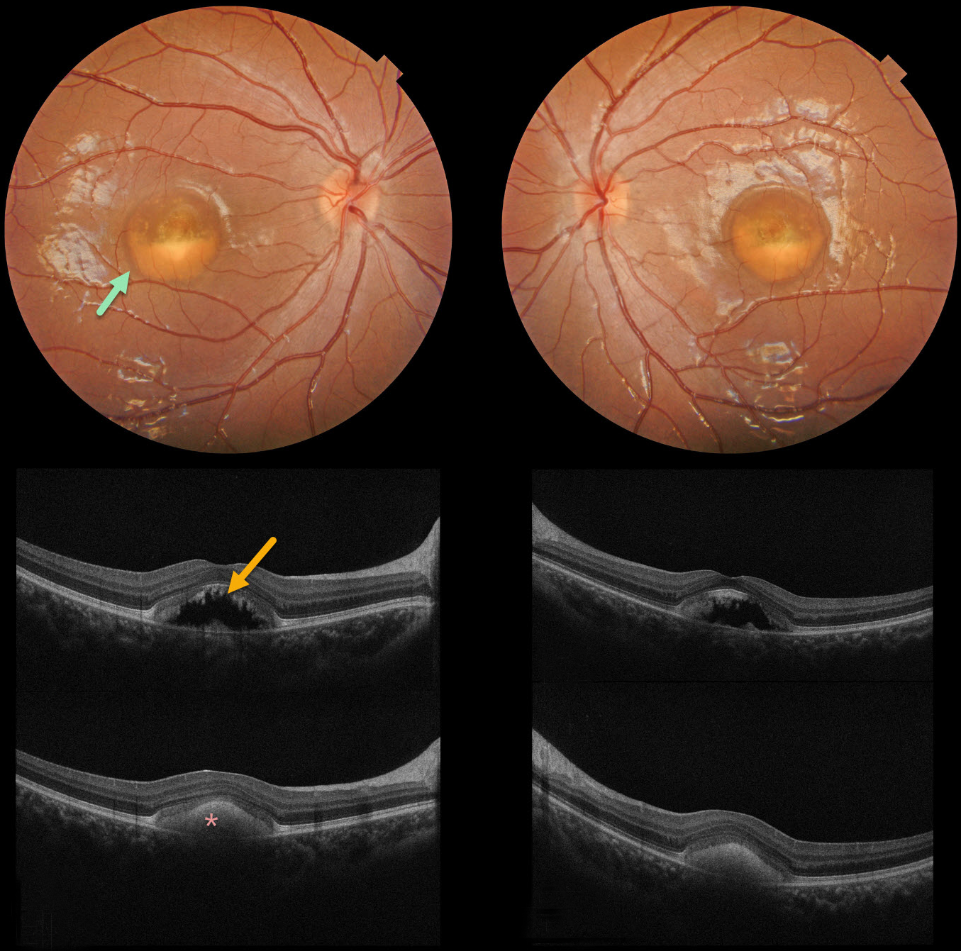
October 2024
BEST DISEASE
Evgenii Chernov
Color photography of our patient’s eyes shows partially scrambled Best lesions. OCT scanning through the superior lesion shows a mostly hyporeflective elevation of the neurosensory retina, with shaggy hyperreflective material along the posterior edge of the outer segments. OCT scanning through the inferior lesion shows hyperreflective layering of the vitelliform material.
Learning Points:
Best disease is associated with a mutation in the BEST1 gene, which encodes bestrophin-1. Bestrophin-1, a calcium-activated chloride channel, is primarily found in the basolateral plasma membrane of the RPE. BEST1 mutations cause a variety of phenotypes, including autosomal recessive bestrophinopathy, best vitelliform macular dystrophy, and autosomal dominant vitreoretinochoroidopathy.
Our patient has progressed to the ‘scrambled egg’ appearance. The remaining vitelliform material gravitates inferiorly, often leaving an optically empty, hyporeflective space between the RPE and outer retina. Eventually, the retina flattens, with secondary outer retinal atrophy.
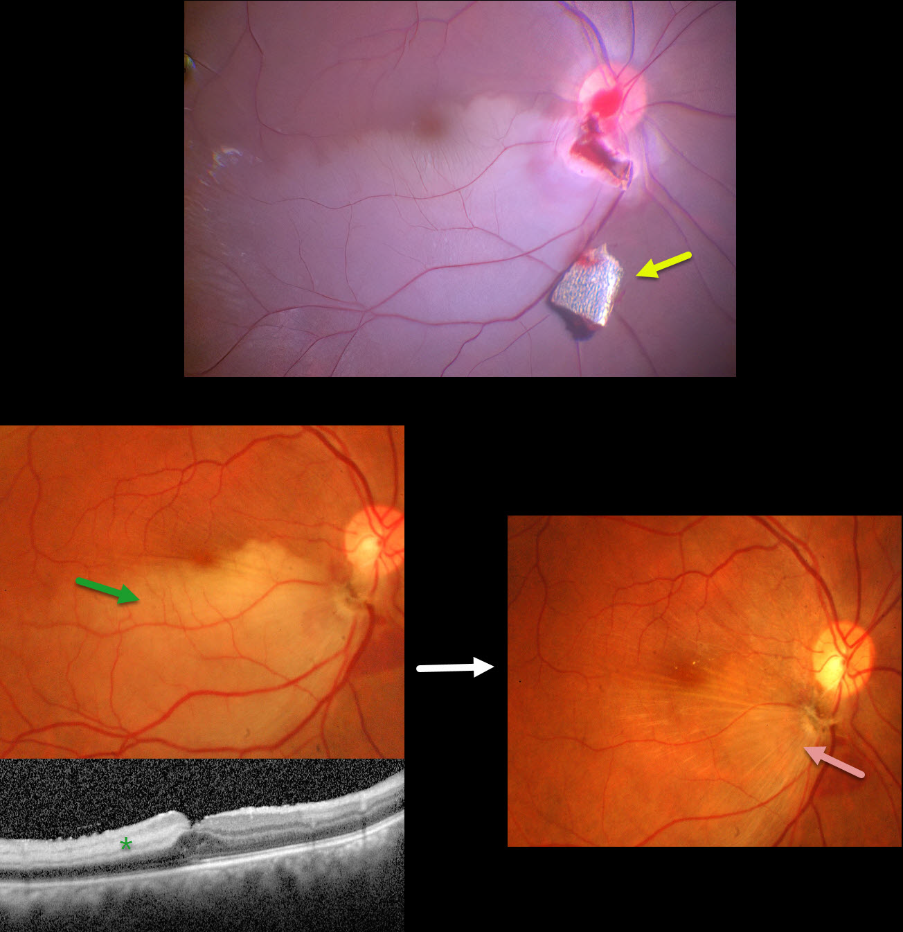
September 2024
INTRAOCULAR FOREIGN BODY WITH SECONDARY BRAO
Anjana Mirajkar and Manish Nagpal
This 34YO male presented with pain and vision loss immediately following a metal-on-metal injury at work. Vision was light perception. Slit lamp examination revealed a corneal laceration and traumatic cataract. B-scan ultrasonography and CT scanning revealed an intraocular foreign body (IOFB). The patient was immediately taken to surgery for corneal wound repair, lensectomy, and vitrectomy.
Intraoperative photography shows a metallic intraocular foreign body (IOFB) resting just inferior to the optic nerve. The impact site is just inferior to the optic nerve, with an inferotemporal branch retinal artery occlusion (BRAO) distally. The IOFB was removed with a rare-earth magnet.
Two weeks following surgery, vision was 20/120 with an aphakic correction. The inferior retina remains opaque, and the OCT shows inner retinal hyperreflectivity from the BRAO. A secondary intraocular lens implantation was performed 3 months postoperatively. Eleven months following the initial surgery, the BRAO is fully resolved, and secondary retinal striae extend outwards from the impact site. Vision remarkably improved to 20/30.
This case was published in the Indian Journal of Ophthalmology (Nagpal M et al, Ind J Ophthalmol 2018;66;146-148).
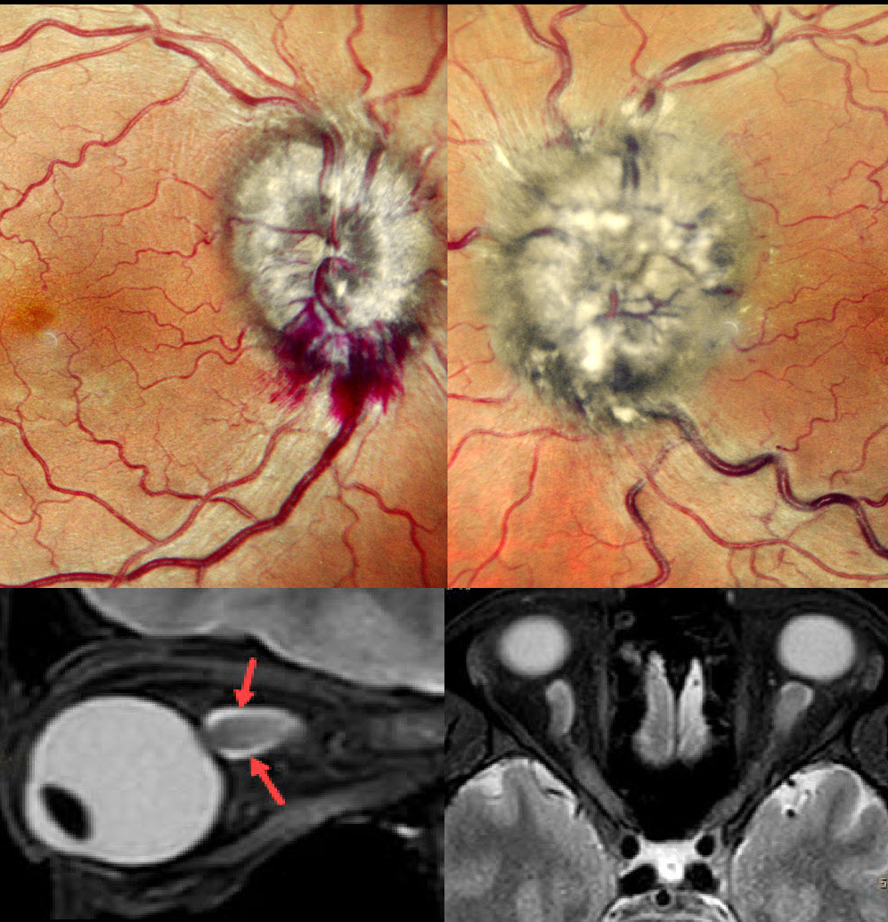
August 2024
LEUKEMIC OPTIC NERVE INFILTRATION WITH PSEUDOPAPILLEDEMA
Anjana Mirajkar and Manish Nagpal
This 40YO male with a known 10-year history of chronic myeloid leukemia complained of vision loss in his left eye for 3 weeks. Vision was 20/20 OD and hand motion OS.
Fundus examination revealed marked bilateral optic nerve swelling, with the left eye greater than the right eye, and some nerve fiber layer hemorrhages along the right inferior disc margin. MRI scanning was consistent with leukemic infiltration of the optic chiasm and optic nerves. She was referred back to her oncologist.
Learning Points:
Leukemia with central nervous system involvement can rarely present with leukemic optic nerve infiltration. This can masquerade as papilledema (bilateral swollen nerves from elevated intracranial pressure) or nerve edema from inflammatory or infectious causes (Miller et al, Graefe’s 2021;259;1315-1322).
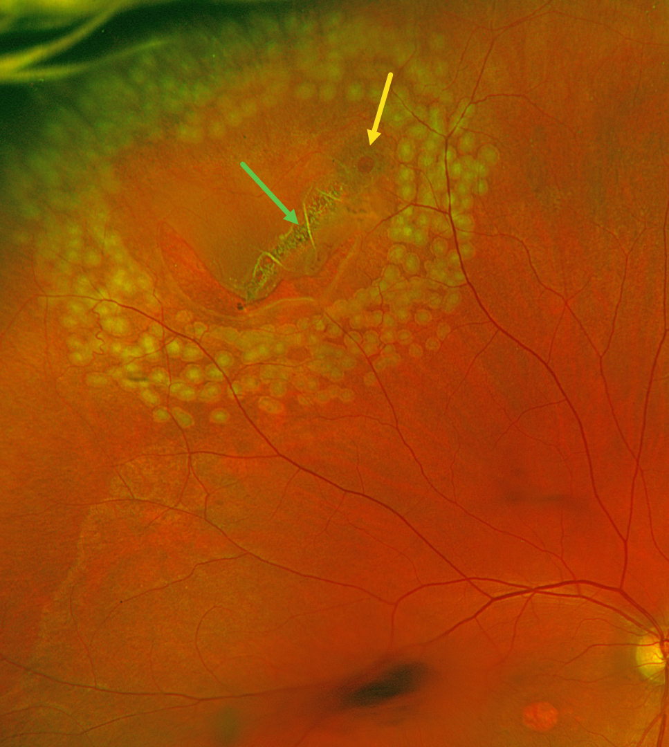
July 2024
RETINAL BREAK
Justin Grassmeyer, Ambar Faridi, and Brittany Heckerman
This 63YO male with history of idiopathic central serous chorioretinopathy (ICSC) with secondary macular neovascularization OD and lattice degeneration OU presented with new floaters in his right eye. He was scheduled for an anti-VEGF injection for the right eye on a treat-and-extend protocol the same day. Vision was stable at 20/100 OD and 20/20 OS. An acute, related retinal tear was found and immediately demarcated with thermal laser photocoagulation.
Optos color RG imaging immediately following the laser procedure shows the retinal break well surrounded by multiple nearly confluent rows of laser burns. The flap of the tear consists of an entire lattice lesion along with a chronic atrophic hole with surrounding pigment. There is an area of central macular atrophy from the ICSC.
Learning Points:
Lattice lesions have a pocket of liquefied vitreous overlying thinned retina with a firm area of surrounding vitreoretinal adhesion. White, fibrosed vessels often crisscross the lesions, which gives the lesions their name, “lattice.” This firm vitreoretinal adhesion, along with the often-present atrophic holes, is what can predispose these eyes to retinal breaks and detachment. Although prophylactic treatment is rarely needed, symptomatic breaks, as in our patient, need prompt retinopexy.
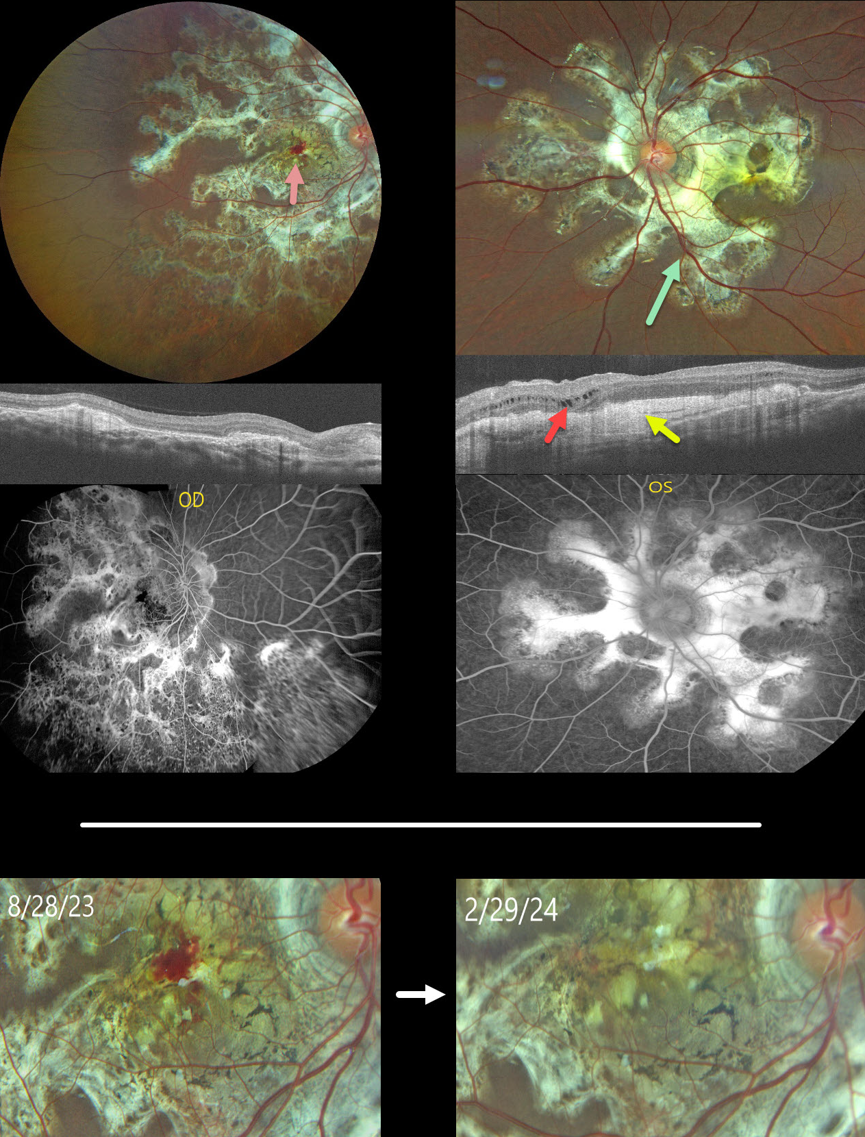
June 2024
TUBERCULAR SERPIGINOUS-LIKE CHORIORETINITIS
Ogugua Okonkwo, Adekunle Olubola Hassan, Ayodele Harriman, Ogochukwu Sibeudu, and Idris Akintayo Oyekunle
This healthy 13YO male presented with a 5-month history of bilateral vision loss. Vision was 20/200 OU.
Fundus photography shows bilateral subretinal, variably fibrotic peripapillary scarring that extends outwards with finger-like projections. A foveal hemorrhage is noted in the right macula. More mottled pigmented scarring extends into the right inferior periphery.
OCT scanning shows variable bilateral outer retinal atrophy and hyperreflective subretinal fibrosis with mild outer nuclear cysts in the nasal left macula. This scarring stains angiographically.
We were hesitant to start anti-VEGF therapy in a young boy and felt that the macular neovascularization causing the right macular blood might be inflammatory. He was therefore placed on a rapidly tapering course of oral prednisone.
At the 6-month follow-up, vision remained stable at 20/200 in this eye, with resolution of the blood.
Mantoux skin testing was negative. We have made multiple attempts to have the patient get QuantiFERON-TB Gold testing, but to date have been unsuccessful.
Learning Points:
Serpiginous choroiditis is an idiopathic, usually bilateral chorioretinal inflammatory condition, most commonly found in middle-aged men (Khanamiri and Rao, Surv Ophthalmology 2013;58:203-232). Recurrent areas of inflammation develop along the edge of a previous scar, producing scarring that meanders from the optic nerve outwards. Vision is good unless scarring or macular neovascularization extends through the macular center.
Tubercular serpiginous-like choroiditis mimics serpiginous but affects younger patients with more multifocal and peripheral recurrences and progression if untreated (Agarwal et al, AJO 2020;220:160-169). Although the exact mechanism is not yet clear, a direct or indirect infectious trigger by the Mycobacterium tuberculosis is thought to cause the choroiditis. We believe our patient has this diagnosis and will continue to confirm it with further testing.
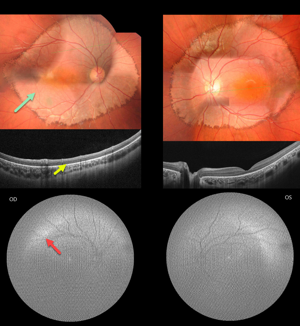
May 2024
INHERITED RETINAL DISEASE
Seif Anwar
This healthy 6YO boy presented with diminished vision noticed by his parents and teacher. There was no family history of eye disease. Vision was 20/200 OU.
Fundus photography shows fairly symmetrical, creamy, pale subretinal lesions encircling each posterior pole. The margins of these lesions have a somewhat irregular hyperpigmented border.
OCT shows outer retinal thinning and loss of the outer retinal band detail, along with a thinned or absent RPE band.
Fundus autofluorescence (FAF) was of extremely poor quality, and possibly shows superior and temporal hyper-FAF just outside each macula. It was quite difficult to obtain imaging in our patient, and these studies required multiple sessions.
Learning Points:
Unfortunately, we do not have a specific diagnosis for our patient’s presumed sporadic inherited retinal disease (IRD). The location and bilaterality of the multimodal findings suggest a cone-mediated disorder. Our patient’s funduscopic appearance somewhat resembles that found in autosomal recessive CDH3 disease, which includes sparse scalp hair and occasional limb abnormalities (Hull et al, JAMA Ophthalmol 2916;134:992-1000). Genetic testing could not be performed due to cost constraints.
Case of the Year
April 2024
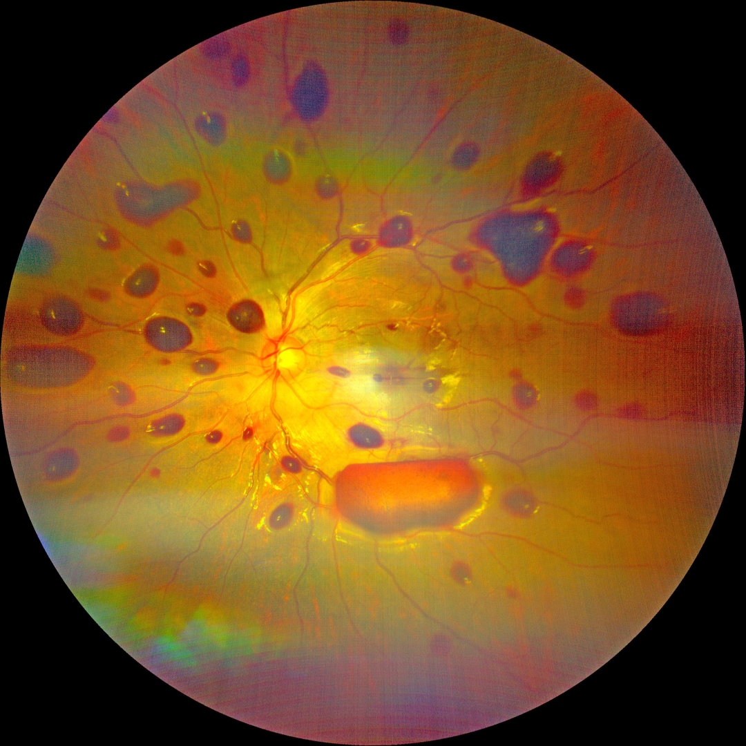
NON-ACCIDENTAL TRAUMA
Kanwaljeet Harjot Madan
This 11YO boy presented with decreased vision in his left eye after his friend’s father thrashed him and was hit in the head 3 days earlier. There was no other sign of bodily injury. Vision was 20/20 in his normal right eye and counting fingers in his left eye.
Our patient’s findings are predominated by numerous mostly sub-ILM hemorrhages. His case is unusual given his age and unilateral findings. Fortunately, with observation, vision recovered to 20/30 three weeks later, with resolution of all findings.
Learning Points:
The shaken baby syndrome is a form of non-accidental traumatic child abuse where the young infant is vigorously shaken, causing whiplash-induced intracranial and intraocular hemorrhaging, often without obvious external trauma.
The ocular findings are usually bilateral, with the hemorrhages often having multiple layers, including subretinal, intraretinal, and preretinal.
March 2024
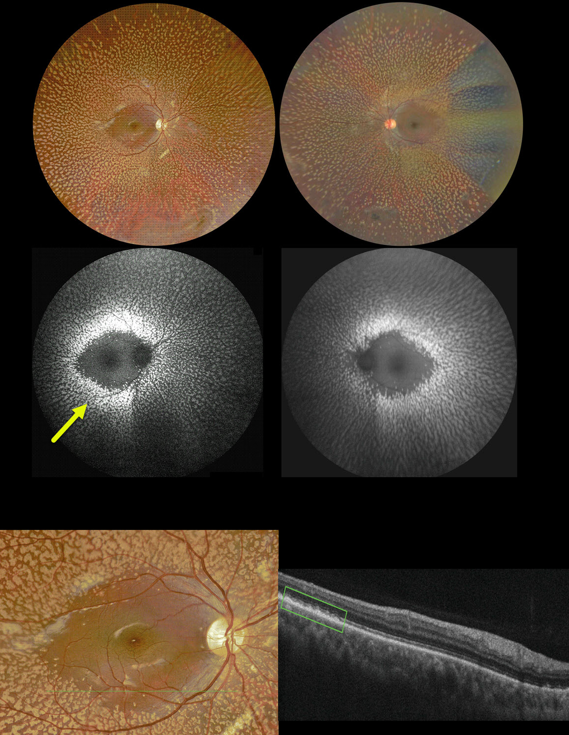
BENIGN FAMILIAL FLECK RETINA
Ayushi Gupta and Vishal Agrawa
This 10YO boy complained of needing frequent changes of his glasses. Vision was 20/20 OD and 20/30 OS. He was a -8D myope OD and -6D myope OS. Corneal topography was consistent with keratoconus.
Color imaging shows symmetric, multiple, discrete yellow-white fleck lesions involving the entire fundus except the maculae. A patch of inferior lattice is also noted OD.
On fundus autofluorescence, these flecks show marked hyper-FAF. On OCT, the flecks appear as hyperreflective outer retinal deposits that extend into and distort the overlying ellipsoid zone.
Learning Points:
Initially reported by Aish and Dajani (BJO 1980;64:652-659), benign familial fleck retina (BFFR) is an extremely rare autosomal recessive disorder that belongs to a heterogeneous group of flecked retina syndromes.
The BFFR gene encodes for a group V phospholipase A2 (PLA2G5). Despite the dramatic fundus appearance, affected individuals have normal acuity, visual fields, and retinal function, including electrophysiology.
Other flecked retinal disorders include fundus albipunctatus, fundus flavimaculatus, familial drusen, retinitis punctata albescens, and fleck retina of Kandori. These disorders can be associated with night blindness, central visual problems, abnormal ERG, and abnormal perimetry.
The ERG helps distinguish BFFR from other flecked retina syndromes, such as retinitis punctata albescens and fundus albipunctatus, as scotopic ERG responses are decreased in the latter conditions but normal in BFFR.
February 2024
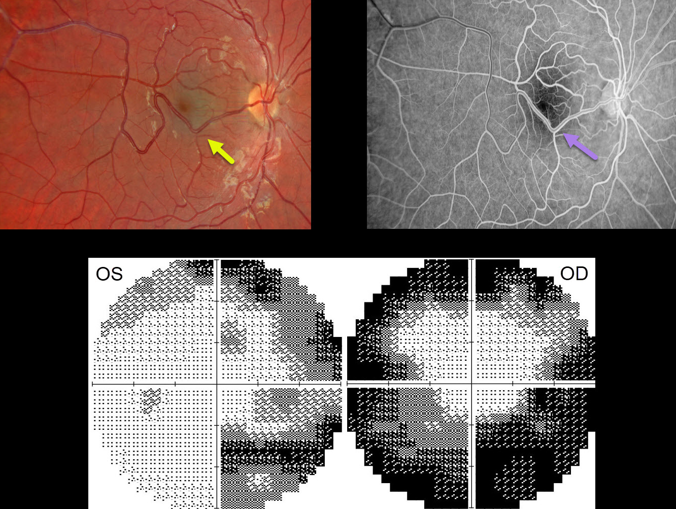
RETINAL VENOUS MALFORMATION
Will Gibson
This 17YO male presented with bilateral vision loss and pain on eye movements 1 week following a COVID-19 infection. There was no prior ocular or medical history. Vision was 20/70 OD and 20/50 OS.
Color imaging of the right eye shows a large temporal retinal venous malformation. This vessel shows angiographic laminar filling without leakage. The right and left nerves were clinically normal, and the left fundus was normal (not shown).
OCT retinal nerve fiber layer analysis was normal bilaterally. Visual fields show moderate generalized constriction OD and nasal loss OS with a possible vertical cut inferiorly. Subsequent neurologic imaging was consistent with optic neuritis. Additional workup was unremarkable, and neuro-ophthalmic consultation led to the diagnosis of bilateral retrobulbar optic neuritis secondary to COVID-19.
Observation was recommended, and symptoms began to improve within a week. Two months later, all visual symptoms resolved, vision returned to 20/25 OU, and the visual fields normalized.
Learning Points:
Retinal vessels virtually always respect the horizontal meridian. Retinal venous malformations are congenital anomalous vessels that, by definition, cross the horizontal.
Originally described by Gary Brown et al as congenital retinal macrovessels (Arch Ophthalmology 1982;100:1430-1436), these vessels are usually located in the macular region and are virtually always venous.
These lesions are now called retinal venous malformations, since about one-quarter of patients have associated venous anomalies in the brain (Pichi et al, JAMA Ophthalmol 2018;136:372-379).
December 2023
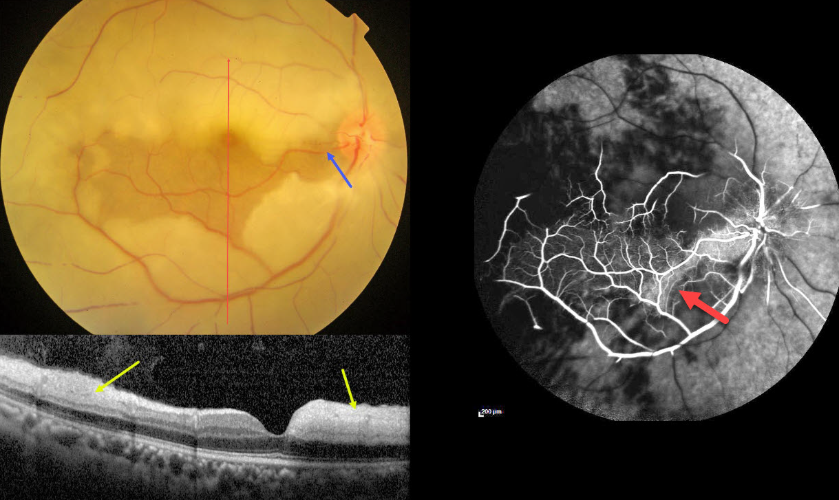
CRAO WITH CILIORETINAL ARTERY SPARING
Anjana Mirajkar and Manish Nagpal
This 45YO male presented with acute vision loss in his right eye 4-5 days earlier. Vision was 20/80 in his right eye and 20/20 in his normal left eye.
Color photography shows an acute central retinal artery occlusion (CRAO) with sparing of the central and inferior macula due to a large cilioretinal artery.
The vertical OCT B-scan shows opaque hyperreflective paracentral inner retina with normal central and inferior foveal layers. Fluorescein angiography confirms preserved central and inferior macular arterial flow.
Workup elsewhere included a normal brain MRI and cardiac 2D Echo. Carotid Doppler showed a plaque in the right internal carotid artery.
Learning Points:
The inner two-thirds of the neurosensory retina is supplied by the central retinal artery, with the choroid supplying the RPE and photoreceptor layer. Acute CRAO, therefore, presents with a white edematous inner retina that spares the fovea.
Cilioretinal arteries, which arise from the posterior choroidal circulation, are present in up to 50% of individuals, and bilateral in about 25% (Schneider et al, Acta Ophthalmologica 2021;99:e310-e318). Our patient was quite fortunate to have a large cilioretinal vessel that relatively preserved central vision.
Patients with acute retinal ischemia (defined as transient monocular vision loss, acute BRAO, or acute CRAO) need to be emergently referred to a stroke center. This is especially urgent with an acute CRAO since about 75% of patients have already developed a recent stroke.
If the patient presents immediately after occlusion, measures to acutely lower intraocular pressure to move the causative embolus downstream can be performed, including ocular massage, paracentesis, and breathing into a bag. Unfortunately, most patients present outside this window (Shah et al, Ophthalmology Retina 2023;7:527-531), and even then, these measures are usually ineffective.
November 2023
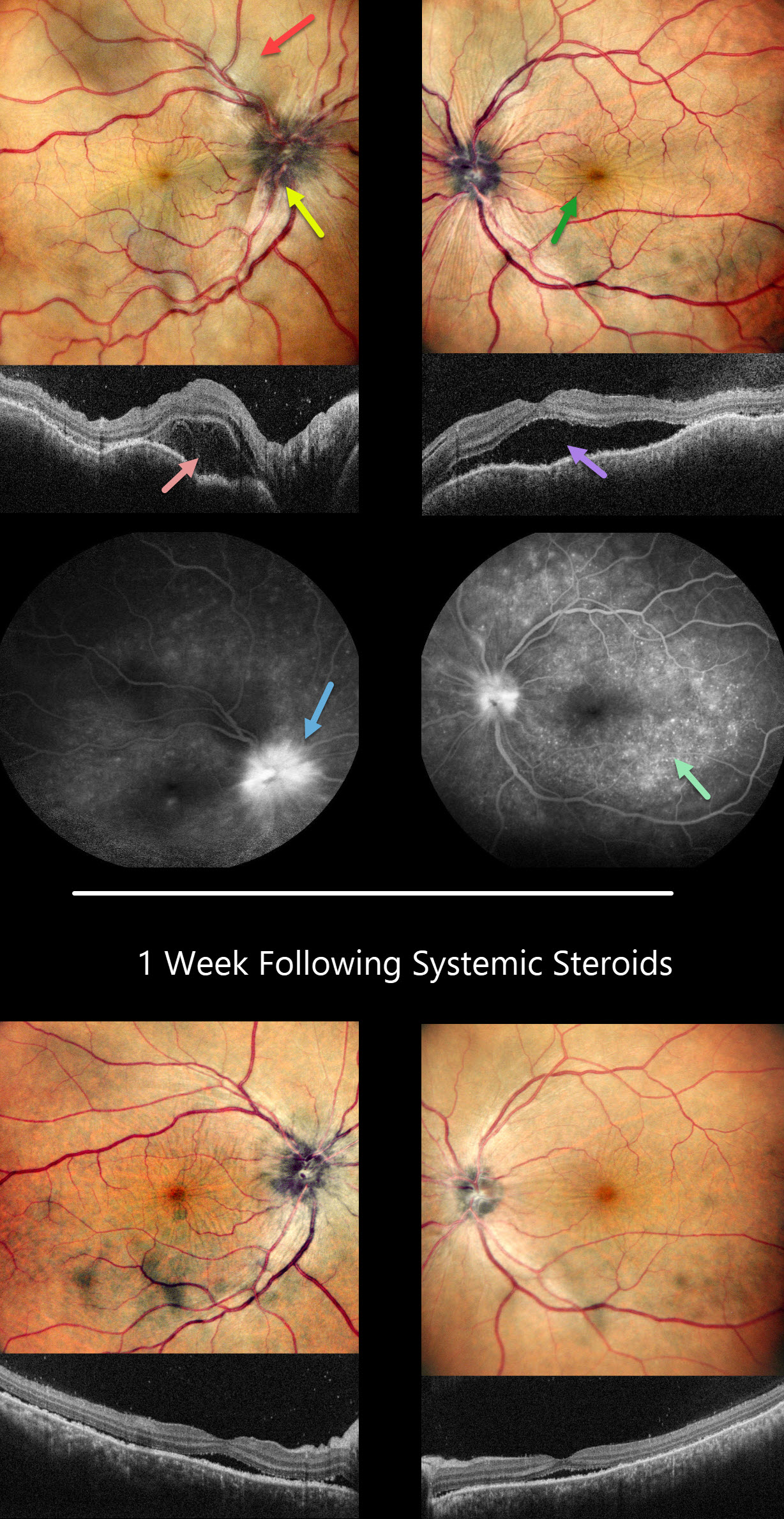
HARADA DISEASE
Anjana Mirajkar, Manish Nagpal and Navneet Mehrotra
This 41YO female presented with headaches, redness, and pain OD for 1.5 months and OS for 2 weeks. She was diagnosed elsewhere with angle closure glaucoma, received a YAG peripheral iridotomy, and started on glaucoma drops. She then saw multiple other doctors who diagnosed her with papilledema.
Her CSF opening pressure was 21, and she was started on oral acetazolamide for a presumed diagnosis of idiopathic intracranial hypertension (IIH). In our office, vision was 20/100 OD and 20/60 OS. Intraocular pressure (IOP) was 30mmHG.
Anterior segments showed shallow anterior chambers with retrolental vitreous cells. Multicolor imaging shows bilateral irregular chorioretinal folds, multifocal serous detachments, and disc hyperemia.
OCT scanning shows a bacillary layer detachment OD, subretinal fluid OS, and a bilateral undulating thickened choroid. Fluorescein angiography shows bilateral optic nerve involvement and pinpoint subretinal leakage.
She was diagnosed with Harada’s disease and started on intravenous methylprednisolone for 3 days, followed by 60mg prednisone PO daily. She was also referred to rheumatology to start immunosuppressants. The acetazolamide was tapered and discontinued for the misdiagnosed IIH.
One week later, vision improved to 20/40 OU. IOP was 10mmHG OU with marked bilateral improvement in the chorioretinal folds, macular fluid, and choroidal thickening. A slow steroid taper was initiated pending initiation of immunosuppressants.
Learning Points:
This case has many classic findings for Harada’s disease, including bilateral panuveitis, optic nerve swelling, thickened choroid, chorioretinal folds, and multifocal serous exudative retinal detachments.
Patients with just ocular findings have Harada’s disease, whereas those with additional systemic findings (including vitiligo, poliosis, headache, vertigo, and hearing loss) have Vogt-Koyanagi-Harada (VKH) disease.
This case also reminds us that it is easy to misdiagnose these complex uveitis cases, especially when not all ocular findings are taken into full consideration in an attempt to give the patient a single, unifying diagnosis.
October 2023
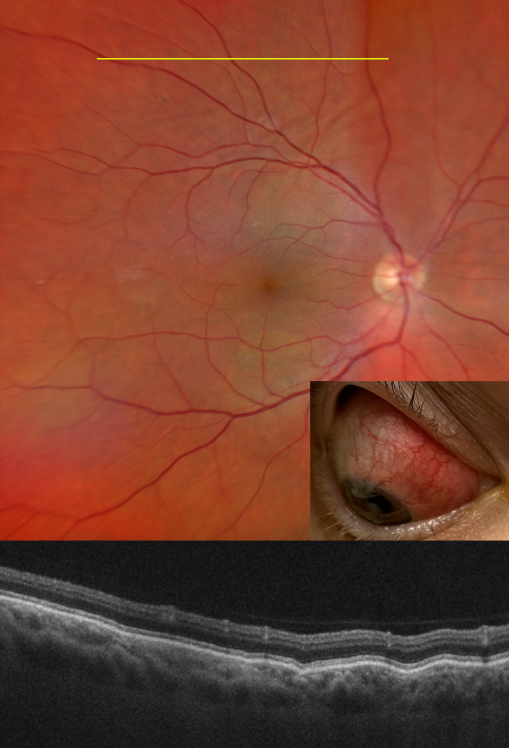
POSTERIOR SCLERITIS
Will Gibson
This 48YO female with a history of psoriatic arthritis presented on 10/25/22 with painful anterior scleritis. Vision was 20/20. The patient was started on 50mg of prednisone and slowly tapered off by rheumatology.
She returned on 7/19/23 with pain behind her right eye. Vision was 20/20. Color imaging shows subtle chorioretinal folds in the superior midperiphery.
OCT B-scan through this region confirmed the chorioretinal folds. B-scan ultrasonography revealed bilateral T signs.
Learning Points:
Posterior scleritis can present either as a diffuse thickening of the entire posterior sclera or as a nodular type with localized scleral thickening (see Agrawal et al, Retina 2016;36:392-401).
Nodular inflammation appears as a posterior pole subretinal yellowish mass lesion, often with overlying serous fluid and chorioretinal folds. B-scan ultrasonography may show fluid in the Tenon space (“T” sign).
Unlike anterior scleritis, the eyes are usually quiet and pain-free. Initial treatment is usually with oral non-steroidal anti-inflammatories or corticosteroids, although patients may also require immunosuppressive therapy. For a large case series of posterior scleritis, see McCluskey et al, Ophthalmology 1999;106:2380-2386.
Oral prednisone was restarted and slowly tapered. Further labs and chest X-ray were performed to rule out other possible causes of scleritis, and she was started on Humira.
September 2023
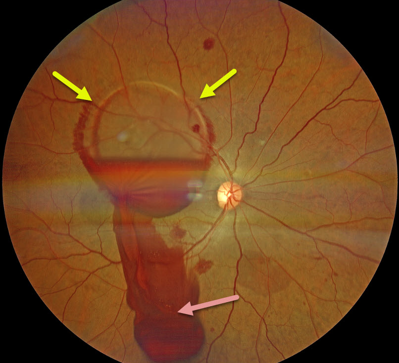
VALSALVA RETINOPATHY
Shivraj Tagare and Nishant Maindargi
This 37YO male presented 1 day after sudden vision loss in his right eye following vigorous coughing. Vision was 20/400 in his right eye and 20/20 in his normal left eye.
Fundus examination of his right eye showed scattered posterior pole retinal hemorrhages and a large sub-internal limiting membrane (ILM) hemorrhage obscuring his right macula.
A Neodymium:YAG laser was used to create a small inferior opening in the ILM. Fundus photography shows that the blood immediately began draining into the inferior vitreous.
Learning Points:
Valsalva retinopathy is characterized by usually unilateral retinal and preretinal hemorrhages caused by raised intrathoracic or intra-abdominal pressure. Various causes include coughing, heavy lifting, and vomiting.
These hemorrhages virtually always resolve without sequelae, although the sub-ILM blood in severe cases can be drained into the inferior vitreous by creating a small ILM opening using either a thermal or Nd:YAG laser, as was done for this patient.
August 2023
RETINAL BREAK
Mohammad Abbas
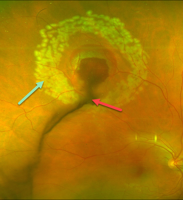
This 27 YO technician working without eye protection in a dermatology laser clinic presented with sudden, painless vision loss and floaters in his right eye. Vision was 20/40.
Optos color imaging shows vitreous blood emanating from the site of laser injury in the super midperiphery. A rim of subretinal blood surrounds the lesion. Fresh laser retinopexy encircles the hemorrhage.
A retinal break is likely obscured by the blood, and this laser should hopefully reduce the risk of a retinal detachment.
Learning Points:
Ocular damage from these lasers depends highly on the laser wavelength, power, and duration. There are several types of lasers used in dermatology, and eye protection for both patients and clinic staff should be used.
June 2023
WAARDENBURG SYNDROME
Gil Calvão-Santos and Keissy Sousa
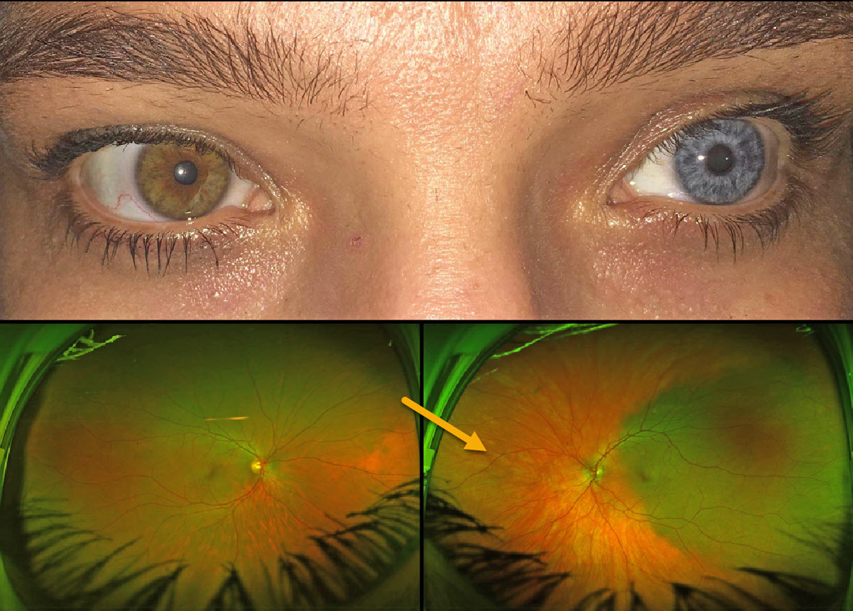
This 43YO female has a constellation of findings that are pathognomonic for Waardenburg syndrome type 1. Vision was 20/20 OU.
External examination shows telecanthus with iris heterochromia. When questioned, she mentioned synophrys (fusion of the eyebrows), which she removed with a laser, poliosis, which she dyed, and a family history of poliosis and heterochromia. She is also congenitally deaf.
Optos imaging shows apparent choroidal melanocytosis temporally in her left eye. But in actuality, this is the only normally pigmented choroid in each eye, with diffuse choroidal hypopigmentation elsewhere.
Learning Points:
Waardenburg syndrome is a group of usually autosomal dominant genetic disorders characterized by achromia of the hair and/or skin, congenital deafness, partial or total iris heterochromia, synophrys, broad and high nasal root, telecanthus, and choroidal hypopigmentation. Vision is usually normal, as was the case in our patient.
See Shields at al for a review of the iris and choroidal abnormalities found in Waardenburg syndrome (JAMA Ophthalmol 2013;131:1167-1173).
July 2023
BEAR TRACKS
Joe Yuenpang Cheung
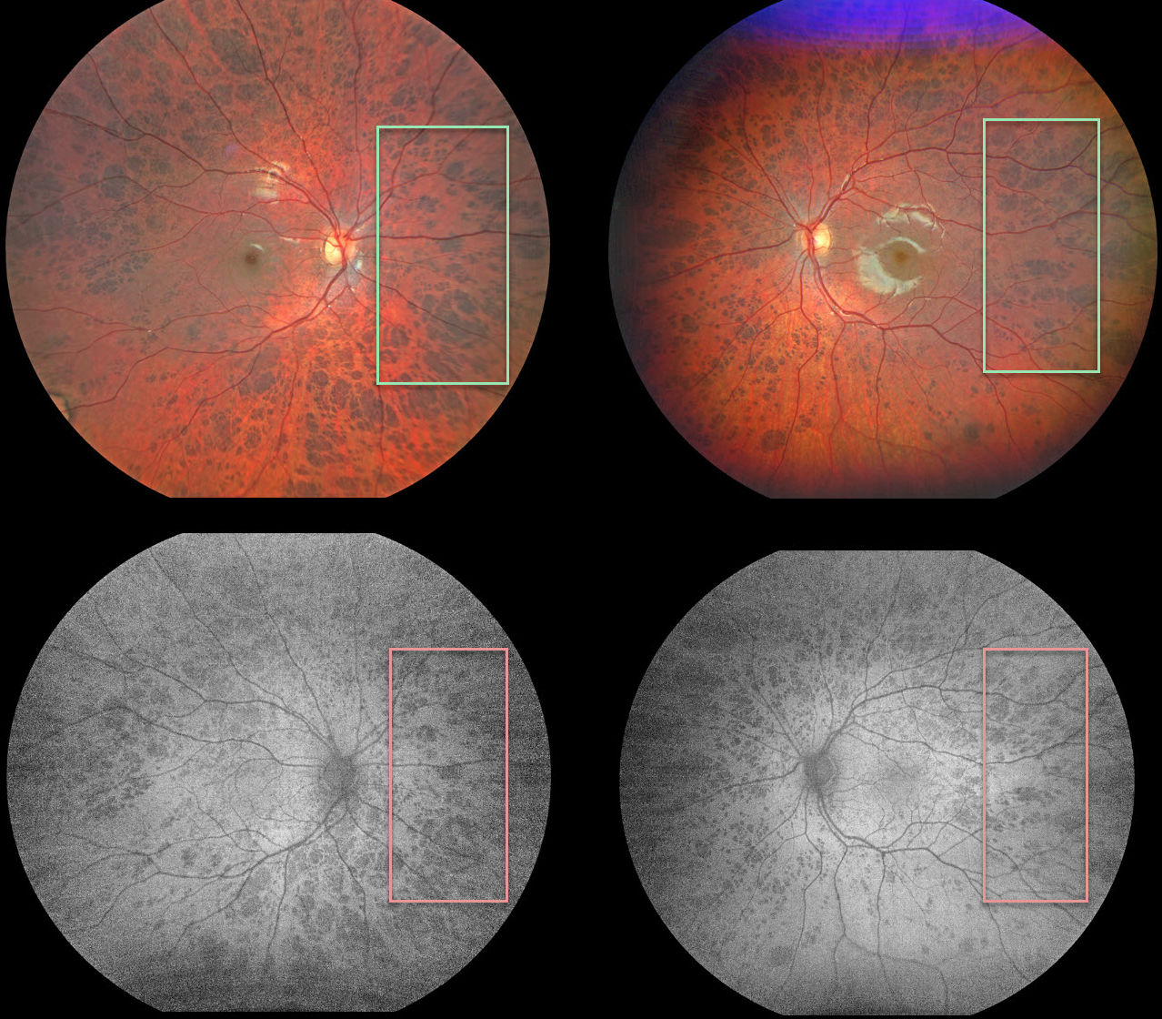
This healthy 8YO was seen for a routine eye examination. Vision was 20/50 OU. Fundus imaging shows extensive bilateral bear track lesions, which are hypoautofluorescent.
Learning Points:
Group-type congenital pigmented nevi of the RPE (bear tracks) is a relatively rare congenital condition characterized by well-demarcated, hyperpigmented, flat, variably sized RPE lesions that resemble bear footprints.
Histopathologically, these lesions are similar to congenital hypertrophy of the RPE (CHRPE), with increased numbers of pigment granules in normal-sized RPE cells. These lesions are benign, cause no visual symptoms, and are not associated with familial polyposis.
The major fluorophore with fundus autofluorescence (FAF) is lipofuscin, which is intraliposomal RPE material generated as byproducts of outer segment metabolism. The outer retina overlying these bear track lesions shows varying degrees of atrophy. This leads to less lipofuscin production with hypo-FAF.
Case of the Year
April 2023
WIDEFIELD OCT
Veronika Matello and Barbara Parolini
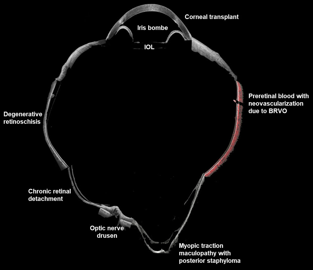
This composite widefield OCT image combines multiple pathologies to create a quite sick virtual eye. All the scans were taken with the Canon Xephilio S1.
Case of the Year
March 2023
RETINAL ARTERIOVENOUS MALFORMATION
Şengül Özdek and Ece Özdemir Zeydanl
This healthy 11YO girl was seen for a routine ophthalmological examination. Vision was 20/20 OU.
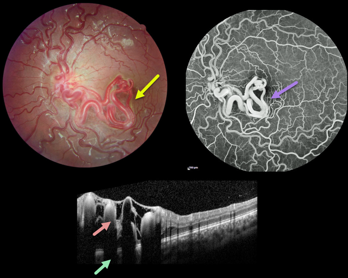
Fundus photography of her left eye shows unilateral tangles of markedly dilated and tortuous vessels extending from the disc into the inferior macula and fovea. The retinal vessels elsewhere are also somewhat dilated and tortuous. OCT scanning through the inferior macula shows these hyperreflective vessels to variably extend through the full-thickness retina with posterior shadowing.
The early-phase fluorescein angiogram shows rapid filling of the dilated artery and vein, with no intervening capillaries (not shown). The late phase shows no angiographic leakage.
Learning Points:
A retinal arteriovenous malformation (AVM) is a congenital connection between an artery and vein without an intervening capillary network. These lesions appear along a spectrum ranging from small isolated lesions to the large ‘bag of worms’ appearance as seen in our patient.
These more extensive lesions are usually part of the Wyburn-Mason Syndrome, which includes vascular lesions in the eyes and midbrain. Retinal venous malformations, also known as retinal macrovessels, are now thought to represent an AVM (Pichi et al., JAMA Ophthalmol 2018;136:372-379).
Fortunately, our patient’s MRI was normal. The AVM should likely remain stable throughout life and should not require any treatment.
March 2023
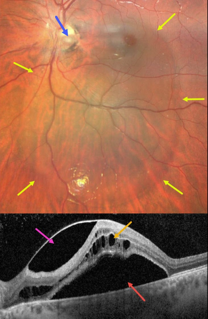
OPTIC NERVE PIT
Emma Oreskovic and Natasha Draca
This 14YO boy presented with these asymptomatic and unilateral findings in his left eye. Vision was 20/50.
Fundus imaging shows a large vertical oval of retinal elevation extending from the superior macula into the inferior midperiphery. This fluid connects to an inferotemporal optic nerve pit.
OCT scanning shows subretinal fluid, cystic fluid mostly in the outer nuclear layer, and fluid below the internal limiting membrane vs within a split nerve fiber layer. He was referred to a retinal specialist for further evaluation and possible treatment.
Learning Points:
An optic nerve pit is a rare (about 0.1% prevalence) congenital anomaly thought to be caused by incomplete closure of the optic fissure during gestation. Pits are most commonly located within or along the inferior-temporal disc margin and usually appear as a gray depression.
While most optic pits are asymptomatic (as in this case), they can cause vision loss by forcing fluid into the inner retina, outer retina, and, ultimately, the subretinal space (optic pit maculopathy). The source of the fluid remains a subject of ongoing debate, with opinions ranging from the vitreous to the subarachnoid space.
A host of treatments for optic pit maculopathy have been suggested over the years. However, a recent meta-analysis suggested that a plain-vanilla vitrectomy without gas tamponade is as successful as other procedures (Zheng et al., Ophthalmology Retina 2020;4:289-299).
February 2023
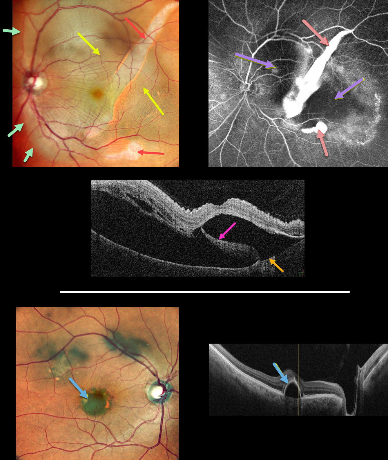
GIANT RPE TEAR
Anjana Mirajkar, Manish Nagpal, Navneet Mehrotr, and Akansha Sharma
This 62YO male presented with 1 month of vision loss in his left eye. Vision was 20/30 OD and 20/60 OS.
Multicolor imaging of his left eye shows a giant RPE tear extending from the inferior macula into the superotemporal midperiphery, along with a much smaller tear just inferior to the macula. The beds of the RPE tears are depigmented. Increased subretinal pigmentation is noted on either edge of the giant tear. Subretinal fluid variably elevates the macula with inner retinal folds radiating from the optic nerve temporally. This subretinal fluid extends into the superior and temporal midperipheries.
B-scan OCT shows marked subretinal fluid. Bare Bruch’s membrane is noted temporally, and the RPE tear is lifted nasally and floating within the serous detachment.
Fluorescein angiography shows marked hyperfluorescent window defects from the absent RPE with blockage on either side of the giant tear from the redundant retracted RPE. He was placed on a 5-day tapering course of oral steroids but was subsequently lost to follow-up.
An asymptomatic retinal pigment epithelial detachment (PED) is noted in his right macula. The choroid does not appear thickened on OCT B-scan.
Learning Points:
A tear of the RPE is most commonly seen in RPE detachments (PEDs) associated with neovascular AMD, but can also occur in numerous other etiologies, including central serous chorioretinopathy and polypoidal choroidal vasculopathy. The tear leaves a depigmented region where the RPE was and is now absent, along with a pigmented, relatively straight subretinal lesion from the scrolled RPE edge.
Our patient’s presentation is unique in that RPE tears are usually much smaller and tend to rip in just one direction. The edges of our patient’s RPE retracted both nasally and temporally. RPE tears are usually singular, and ours had an additional, much smaller secondary tear more inferiorly.
Although our patient’s findings seem to be in the pachychoroid spectrum, the lack of a thickened choroid on OCT scanning makes the etiology less certain.
See Ersoz et al for a great review on RPE tear classification, pathogenesis, predictors and management (Surv Ophthalmology 2017;62:493-505).
November 2022
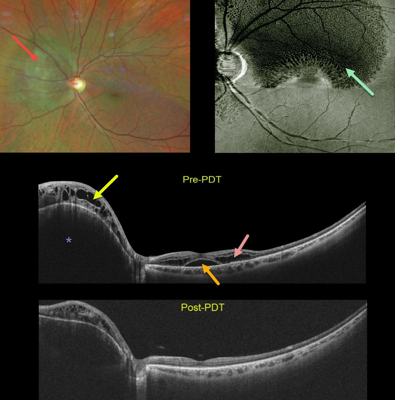
CHOROIDAL HEMANGIOMA
Barbara Parolini and Veronika Matello
This healthy 45YO female was referred for macular schisis. Vision was 20/50. Color photography shows an orange-red subretinal lesion nasal to the nerve.
Widefield OCT shows an elevated hyporeflective choroidal mass with overlying cystic retinal edema. Subfoveal fluid with cystic fluid in the outer retina is noted in the macula. An en face OCT of the outer retina more dramatically shows the cystic changes.
The choroidal lesion with overlying retinal edema dramatically improved following photodynamic therapy. Vision improved to 20/20.
Learning Points:
Widefield imaging is revolutionizing the way we diagnose and treat retinal disease. This case is a perfect example in which macular imaging alone could lead to an incorrect diagnosis.
In this case, the macular findings in isolation suggest either optic pit maculopathy (Imamura et al, Retina 2010;30:1104-1112) or paraproteinemic maculopathy (Mansour et al, Ophthalmology 2014;121:1925-1932). This widefield technology led to a correct diagnosis, allowing for appropriate treatment with excellent anatomic and visual results.
August 2022
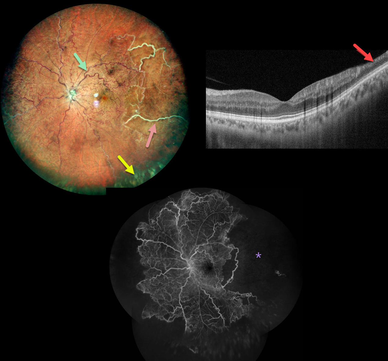
ISCHEMIC CRVO
Nivesh Gupta
This healthy 34YO male presented with this relatively asymptomatic central retinal vein occlusion (CRVO). Vision was 20/20 OU.
Color imaging shows large temporal nonperfused retinal vessels with old inferior yellow vitreous hemorrhage. The retinal vessels are diffusely dilated and tortuous. OCT B-scan shows a remarkably normal central macula with some distal inner retinal thinning. Fluorescein angiography confirms widespread peripheral nonperfusion.
Anti-VEGF therapy, followed by panretinal photocoagulation, was administered.
Learning Points:
The Central Vein Occlusion Study (CVOS) defined ischemic CRVO as having at least 10 disc areas of angiographic retinal nonperfusion as determined by standard photographic views with the Canon (60 degrees) or Topcon (45 degrees) wide-angle fundus cameras (Ophthalmology 1995;102:1434-1444).
Although clearly ischemic, this patient might have been classified by the CVOS as a perfused occlusion, depending on the extent of peripheral ischemia imaged. As with diabetic retinopathy (see Brucker, Retina 2021;41:461-463), ultrawidefield imaging and anti-VEGF injections have thrown a literal wrench into how we think about, classify, and treat retinal disorders.
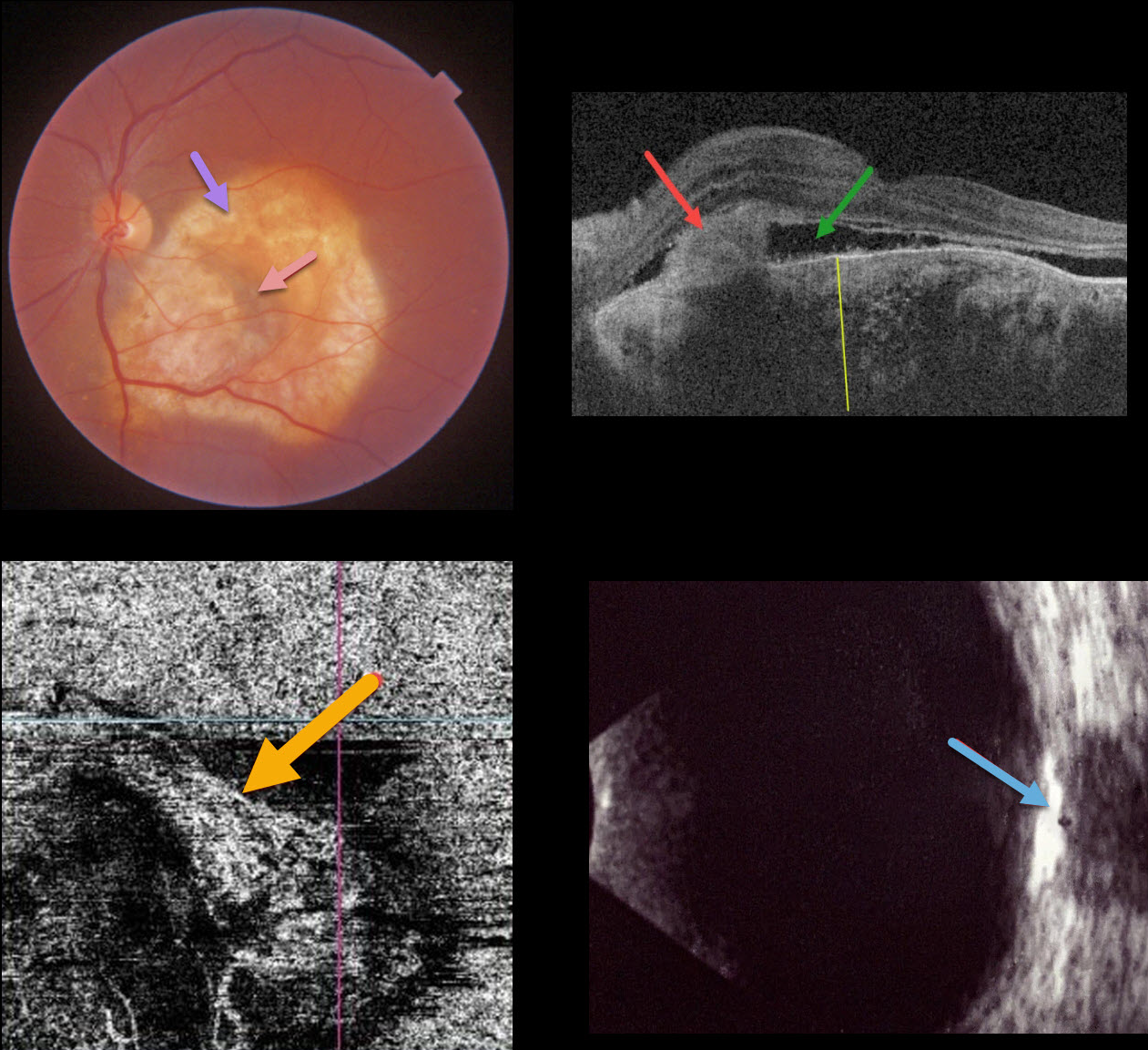
July 2022
CHOROIDAL OSTEOMA
Omar Mulki
This 40YO female presented with recent vision loss in her left eye. Vision was 20/20 in her normal right eye and counting fingers in her left eye.
Triton color imaging shows an amelanotic choroidal lesion occupying virtually the entire macula. There is a small dot of central blood. Swept-source OCT shows a markedly thickened choroid with variable regions of hyper- and hyporeflectivity. A hyperreflective type 2 macular neovascularization (MNV) is seen along with exudative subretinal fluid. OCT angiography shows flow signals within the MNV. B-scan ultrasonography shows the lesion to be markedly hyperreflective.
An anti-VEGF injection was given, but she was unfortunately subsequently lost to follow-up.
Learning Points:
Choroidal osteomas are benign, calcified amelanotic choroidal tumors. They are more common in women, are almost always unilateral, and are usually located in the peripapillary or macular regions.
Choroidal lesions are best seen on the Optos red channel. Fluorescein angiography (FA) usually shows early patchy hyperfluorescence with late diffuse staining. Ultrasonography is often helpful in differentiating an osteoma (markedly hyperreflective) from other amelanotic choroidal tumors.
Macular neovascularization develops in a significant number of eyes and can be successfully treated with anti-VEGF therapy (see Seong et al, Graefe’s 2022;250:1713-1721).
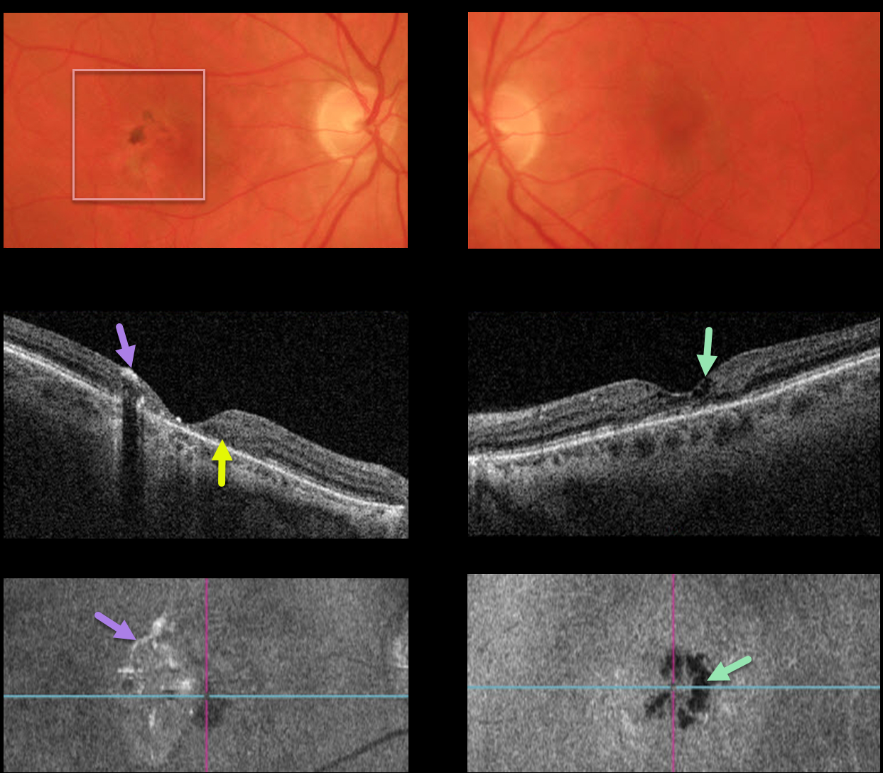
MACULAR TELANGIECTASIA
This 60YO female presented with 20/70 vision OD and 20/50 vision OS, along with classic findings of macular telangiectasia type 2 (MacTel2).
The perifoveal retina is somewhat opaque with significant pigmentary changes, particularly in the right eye.
OCT scanning of the right eye shows significant central outer retinal atrophy with hyper-reflective pigment migration. The left OCT shows milder atrophy, central inner cavitations, but no pigment migration.
En face imaging helps highlight some of the findings described above. The right en face image shows scattered patchy hyperreflective areas corresponding to the pigment migration, while the left en face image highlights the hyporeflective cavitations.
Learning Points:
Originally described by Gass (Arch Ophthalmology 1982;100:769-780), MacTel2 is a neurodegenerative disorder, most likely originating from Müller cell dysfunction. The term telangiectasia is misleading because the funduscopic findings are mostly nonvascular.
The refractile inner retinal ‘crystals’ are thought to be Müller cell footplates. The foveal retina shows a whitish discoloration, most likely due to retinal opacification resulting from Müller cell dysfunction.
Photoreceptor loss allows RPE cells to migrate along intraretinal capillaries, causing the coarse clinical pigment clumping (not seen in our patient).
The majority of patients will also have subclinical chorioretinal anastomosis associated with right-angle venules (Spaide et al, Retina 2018;38:1920-1929).
Secondary macular neovascularization can be successfully treated according to age-related macular degeneration protocols.
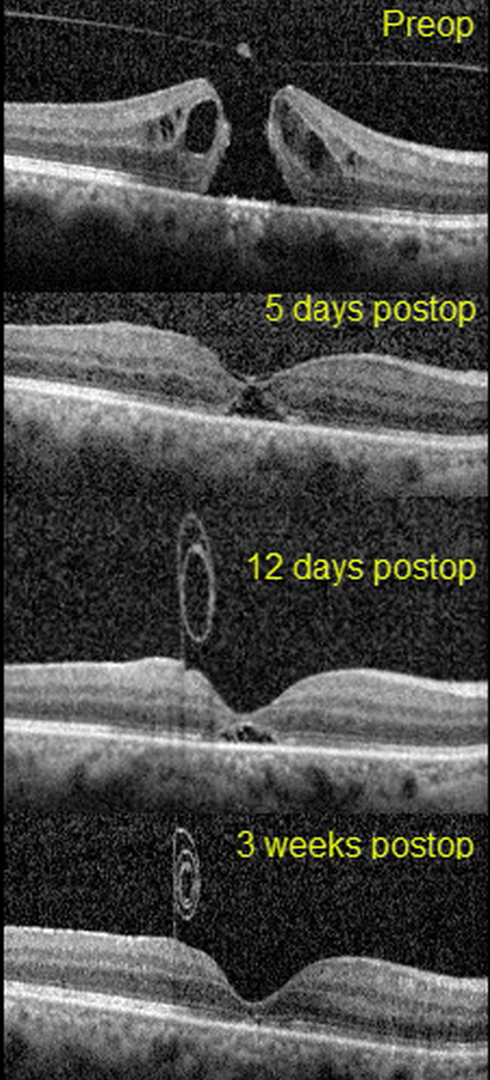
March 2022
MACULAR HOLE
Erdem Dinç
This 51yo presented with 20/1250 vision in her right eye from a primary full-thickness macular hole without traction measuring 407 microns. There was also a moderately severe nuclear sclerotic cataract.
Combined cataract surgery with 23-gauge pars plana vitrectomy, temporal half-moon inverted ILM flap, and fluid-air-SF6 exchange was performed, followed by prone positioning.
On the 5th postoperative day, the flap extended over the closed hole, but one week later, the ILM flap retracted and was rolled inward, suspended anterior to the macula.
Three weeks following vitrectomy, the hole was completely flattened with partial restoration of the outer retinal layers. The ILM flap continued to contract and scroll onto itself. Vision was 20/40.
Learning Points:
Introduced by Kelly and Wendel (Arch Ophthalmol 1991;109:654-659), macular hole surgery is one of the modern success stories in ophthalmology. ILM flaps appear to improve the surgical success for large (>400 microns) macular holes.
In this case, the ILM flap inadvertently scrolled onto itself and away from the macular hole by post-op day 12. Some authors have suggested that the ILM flap provides a scaffold for cells to grow over and help heal the hole.
However, most macular holes are actually foveal dehiscences without tissue loss, where the foveal retina splits and elevates like a drawbridge (Tornambe, Retina 2003;23:421-424). For successful surgery, there is thus no need or significant evidence for glial or neurosensory retina regrowth, just for the “drawbridge” to close.
As this case exemplifies, the ILM flap most likely serves the same purpose as the gas bubble, helping to sequester the macular hole from the overlying vitreous. This allows the RPE to actively pump (and the choroid to passively absorb) the subretinal fluid, allowing the foveal tissue to flatten into its normal position. Once the hole closes, as long as the vitreous and epimacular traction are relieved, there is likely no need for the ILM flap.
Case of the Year
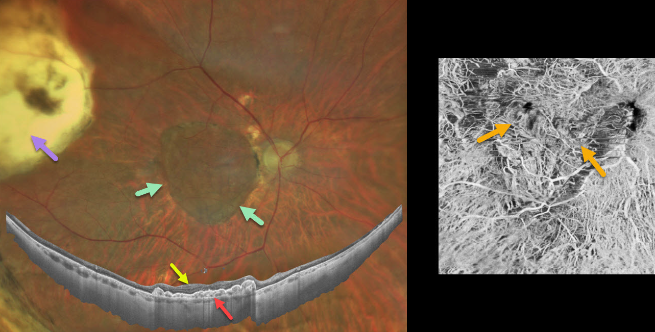
January 2022
CHOROIDAL TRANSPLANT
Barbara Parolini and Veronika Matello
This patient is a teacher who suffered from a wet AMD submacular hemorrhage. Despite six anti-VEGF injections, vision dropped to 20/200. She was 58 years old and unable to read.
After a long discussion regarding the pros vs cons of surgery, she underwent autologous choroidal transplantation in 2013.
The surgical procedure included:
1) Complete pars plana vitrectomy
2) Creation of a temporal retinal detachment
3) Peripheral 200-degree retinotomy
4) Macular neovascularization (MNV) removal
5) Feeder vessel endodiathermy
6) Designing the peripheral choroidal patch with endodiathermy
7) Cutting and isolation of a full-thickness autologous RPE and choroid transplant (under perfluorocarbon liquid, PFCL)
8) Translocation of the patch under the fovea (under PFCL)
9) Transfer of PFCL from under to over the retina with retinal reattachment
10) Peripheral laser
11) PFCL exchange for 1000cs silicone oil.
Eight years later, a Clarus 500 widefield photograph shows the intact submacular choroidal autograft and a white area of bare sclera in the temporal quadrant, which was the harvesting site for the autograft.
Canon Xephilio S1 widefield OCT shows a fairly normal retina overlying the graft. Xephilio A1 angioOCT shows the intact choroidal graft vessels arranged in a different direction compared to the native surrounding choroidal vasculature.
When last examined, near vision was 20/30. She recently developed an MNV in the contralateral eye, which is being treated with anti-VEGF injections.
May 2023
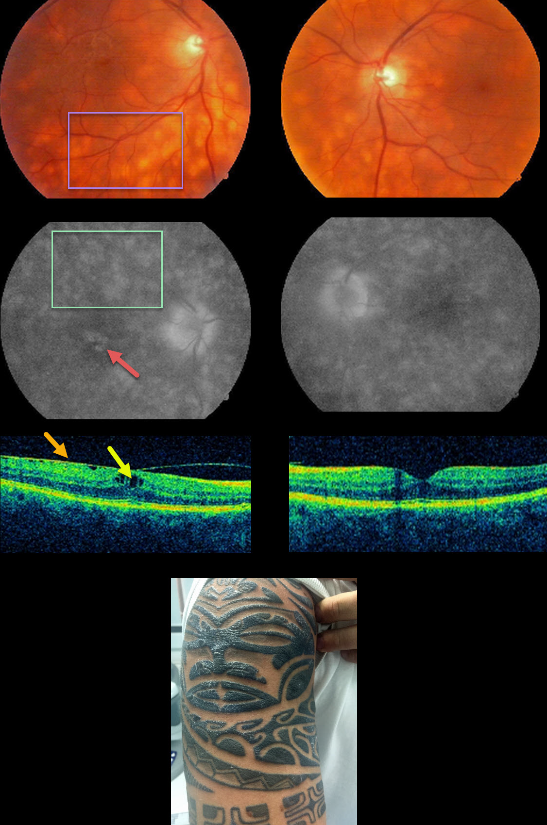
TATTOO-INDUCED UVEITIS
Natasa Draca
This 39YO male presented with bilateral floaters and a several-year history of intermittent panuveitis. He had a history of treated hepatitis C.
Vision was 20/25 OD and 20/20 OS. Anterior segments were normal. There was mild bilateral vitritis and scattered amelanotic subretinal lesions in each posterior pole.
Fluorescein angiography shows staining of these lesions with cystoid leakage in his right macula. OCT scanning shows a mild epiretinal membrane with cystoid edema OD. The initial differential diagnosis included birdshot chorioretinopathy and sarcoidosis.
HLA-A29 was negative, but on further questioning, he told us that the tattoos on his arms always became swollen and painful prior to the recurrent uveitis.
A biopsy of one of the inflamed tattoos revealed inflammation granulomatosa. The granulomas were classified as foreign-body or sarcoid-type reactions.
However, the sarcoidosis workup, including ACE, chest X-ray, and chest CT, was all normal. He is being treated as needed with oral and topical steroids.
Learning Points:
Tattoo pigments are known to produce mast cell activation and to provoke cellular lysis after phagocytosis. Intradermal injection of melanin, interphotoreceptor retinoid-binding protein, and S-antigen is known to produce a granulomatous anterior or pan-uveitis (see Ostheimer et al, AJO 2014;158:637-643). The uveitis can be controlled with systemic steroids and/or immunosuppressants.
December 2021
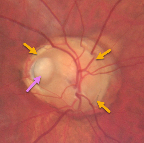
OPTIC NERVE PIT
Otis Hertsenberg
This 36YO male presented with an asymptomatic anomalous right optic nerve coloboma with a large temporal pit.
The superior portion of the nerve is fairly normal. The colobomatous portion of the nerve is enlarged and excavated nasally, inferiorly, and temporally.
Vision was 20/20 OD and 20/400 OS. He had a history of surgery for a congenital cataract and microphthalmia in his left eye.
Learning Points:
Optic pits are most commonly located within or along the inferior-temporal disc margin and usually appear as a gray depression. They are thought to be caused by incomplete closure of the optic fissure during gestation.
While most optic pits are asymptomatic, they can cause vision loss by forcing fluid into the inner retina, outer retina, and, ultimately, the subretinal space (optic pit maculopathy). The source of the fluid remains a subject of ongoing debate, with opinions ranging from the vitreous to the subarachnoid space.
November 2021
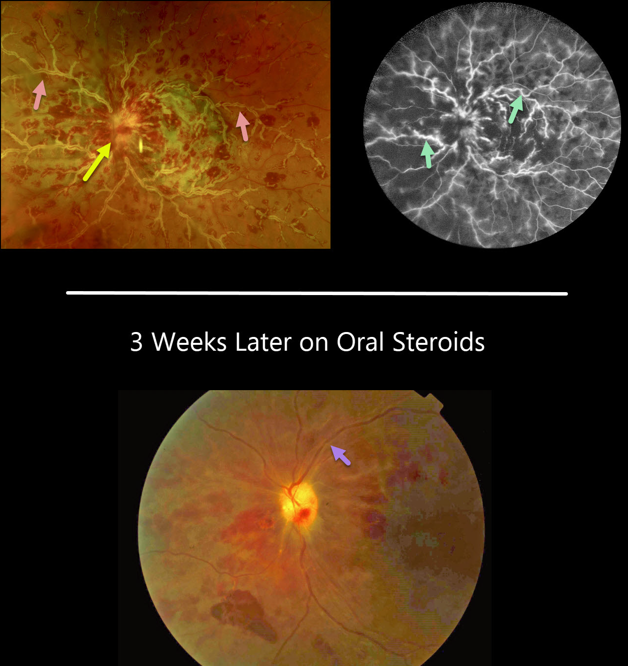
FROSTED BRANCH ANGIITIS FROM SARCOIDOSIS
David Kilpatrick
This 34YO African American female presented with several days of painless vision loss in her left eye. Vision was 20/30 OD and HM OS with an afferent defect OS. Anterior segments were normal.
There was mild disc edema OD (not shown) and florid disc edema OS with retinal vasculitis predominantly involving the veins.
On fluorescein angiography, the retinal veins in the left eye were diffusely stained.
On further review of systems, she had a new skin rash on her right forearm, but was otherwise asymptomatic. Testing revealed elevated angiotensin-converting enzyme and lysozyme levels, and a chest X-ray showed bilateral perihilar fullness.
With a working diagnosis of sarcoidosis, she is currently being managed with oral steroids and has been evaluated by both rheumatology and pulmonology.
While her retinal vasculitis has responded well to treatment, her vision remains HM three weeks later.
Learning Points:
Frosted branch angiitis is a retinal vasculitis that gets its name due to the involved vessels resembling the branches of a tree that are “frosted” with snow. Although these findings may be isolated, they are often considered a phenotype found in a multitude of inflammatory conditions, most commonly CMV retinitis.
The inflammation is usually highly responsive to steroids, and the visual prognosis is generally good, although some may develop secondary neovascularization in the anterior and posterior segments due to widespread ischemia.
October 2021
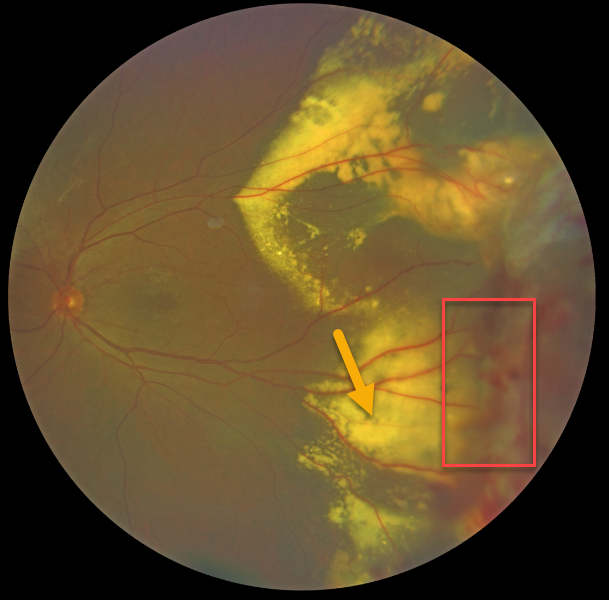
COATS DISEASE
Kumar Chugani
This 66YO male presented with classic Coats disease findings. Vision was 20/30.
Confluent subretinal lipid extends into the peripheral temporal macula. The source for this lipid is noted in the temporal midperiphery, with dilated, irregular telangiectatic vessels with aneurysmal dilations.
Learning Points:
Laser photocoagulation can be used to treat leaking macroaneurysms and telangiectatic vessels in Coats disease. Anti-VEGF injections can also be used to stabilize vascular leakage.


