

Cuticular Drusen
<< Back to Cases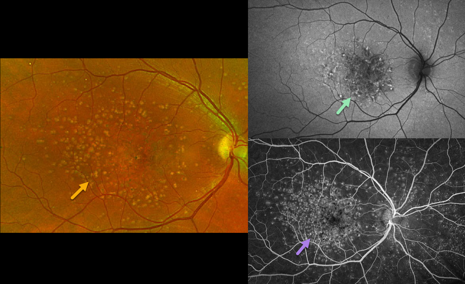
CUTICULAR DRUSEN
Originally posted on @retina.rocks September 25, 2026
This asymptomatic, healthy 45YO male was referred for age-related macular degeneration, which was diagnosed during his 30s. Vision was 20/20 OU.
Optos color RG imaging of his right eye shows mixed drusen with pigment clumps. The drusen on fundus autofluorescence (FAF) are mostly hyper-FAF. Fluorescein angiography reveals innumerable hyperfluorescent drusen. Identical findings were present in his left eye (not shown).
Learning Points:
Originally described by Gass as basal laminar drusen (AJO 1985;99:445-459), cuticular drusen were initially thought to represent nodular thickening of the RPE basement membrane. They appear clinically as numerous, round, yellow-white ‘hard drusen’, and on OCT as small, arrowhead-shaped drusen located above Bruch’s membrane and below the RPE (Spaide and Curcio, Retina 2010;30:1441-1454). Our patient’s angiographic appearance echoes Dr. Gass’ original description of “a beautiful stars-in-the-sky or Milky Way pattern” of hyperfluorescence. Cuticular drusen are classically associated with avascular vitelliform lesions (Finger et al, Retina 2010;30:1455-1464).


