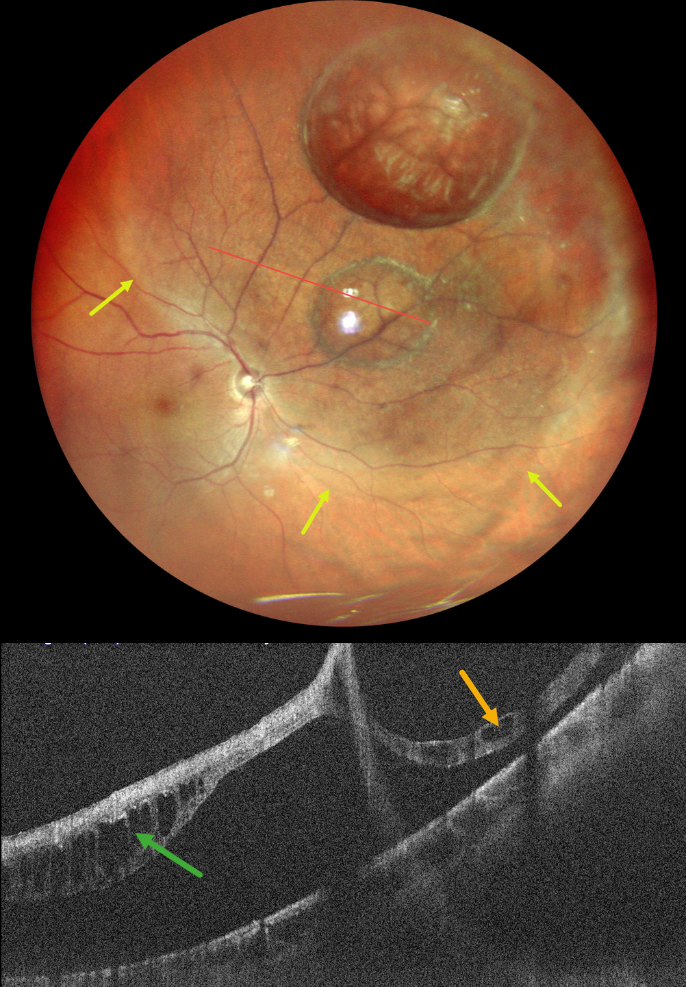
This healthy 43YO male presented with inferotemporal field loss in his right eye for 2 weeks. Vision was 20/20 OU.
Pseudocolor SLO imaging shows a superonasal macula-on rhegmatogenous retinal detachment (RRD) with two prominent retinal cysts.
OCT confirms the retinal detachment with cystic outer retinal fluid within the detachment and within the outer margin of a cyst. Emergent pars plana vitrectomy was scheduled.
Learning Points:
A retinal cyst is defined as a fluid-filled space in or derived from the retina, the diameter of which is greater than the thickness of the normal retina. They most commonly occur in chronic retinal detachments and have historically been described as retinal macrocysts (Liu et al, JAMA Ophthalmology 2018;136:956-958). The lining of these cavities localizes to the outer plexiform layer, indicating that the lesions result from intraretinal splitting rather than representing true cysts. In our experience, these cysts promptly resolve following retinal reattachment and therefore don’t need to be drained directly during retinal reattachment surgery.