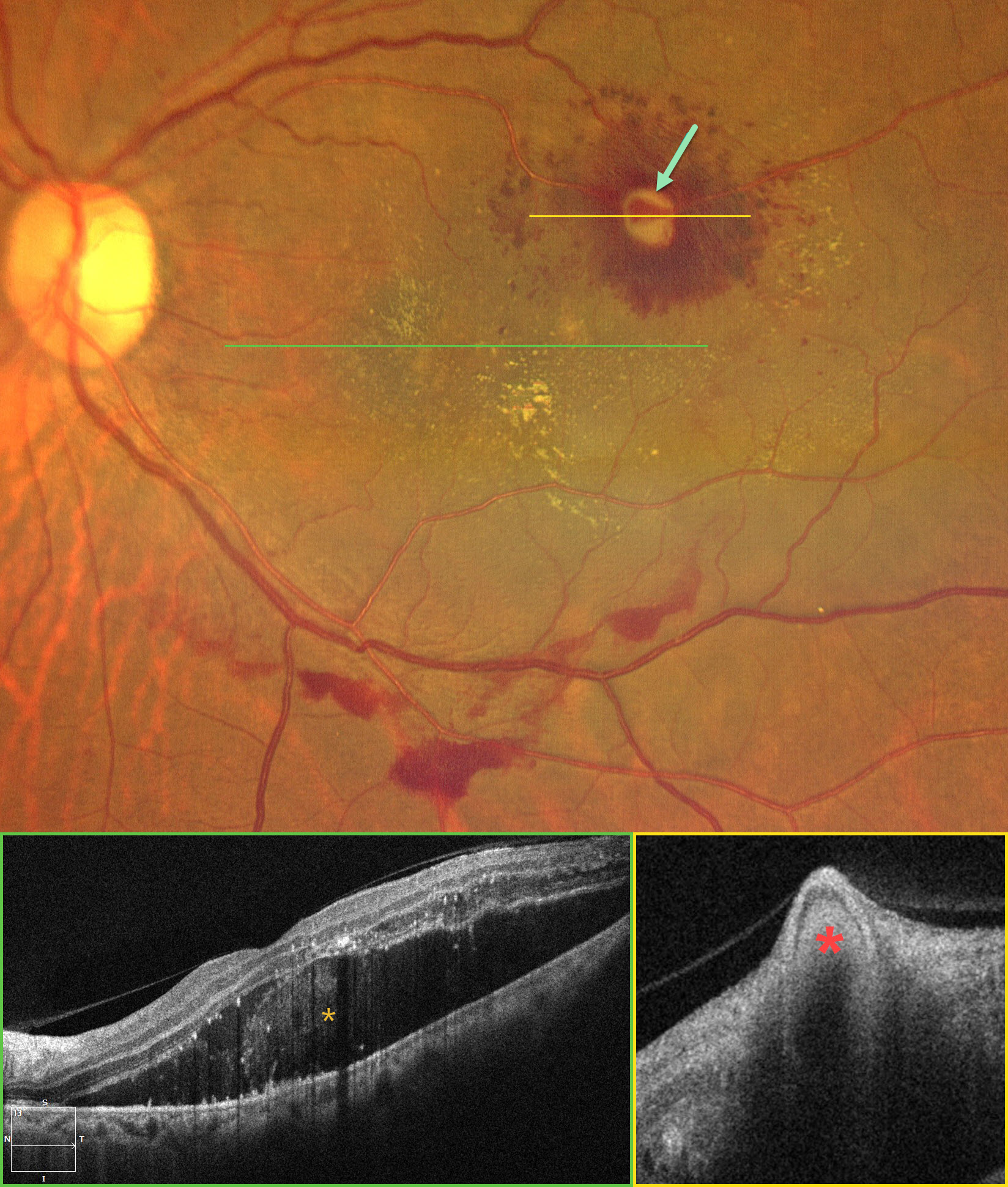
This 62YO female presented with a 1-week history of decreased vision in her left eye. Vision was 20/20 in her normal OD and 20/200 OS.
Color photography shows a fibrosed RAM in the superotemporal macula surrounded by intraretinal hemorrhages. Fine lipid deposits are noted temporal, inferior, and nasal to the RAM. Multiple small patches of preretinal blood are present along the inferotemporal arcade. OCT scanning through the central macula shows a retinal detachment with variable hyperreflective subretinal fluid. Hyperreflective lipid is noted in the mid to outer retina, and hyperreflective subretinal foci also probably represent lipid deposits. The hyper-reflective RAM indents the inner retina.
Learning Points:
A RAM is thought to occur in a weakened arterial wall from arteriosclerosis and appears as a yellow-white dilated lesion along a retinal artery within the first 3 bifurcations from the optic nerve. There are 2 distinct presentations: hemorrhagic or exudative. The hemorrhagic type often presents, as in this case, with pathognomonic multiple layers of blood. Acute macular exudation can be treated with observation, anti-VEGF injections, or thermal laser.
An intravitreal ranibizumab injection was given. Thermal laser to the RAM was planned but unfortunately she was immediately lost to follow-up.