This 76YO female has been followed by us for several years with recurrent anterior scleritis. Her personal medical history on presentation was negative for established underlying rheumatic disease. She presented for a regular follow-up examination with symptoms of mild pain OD. Vision was 20/30 OD and 20/30 in her normal OS. Anterior segments were quiet.
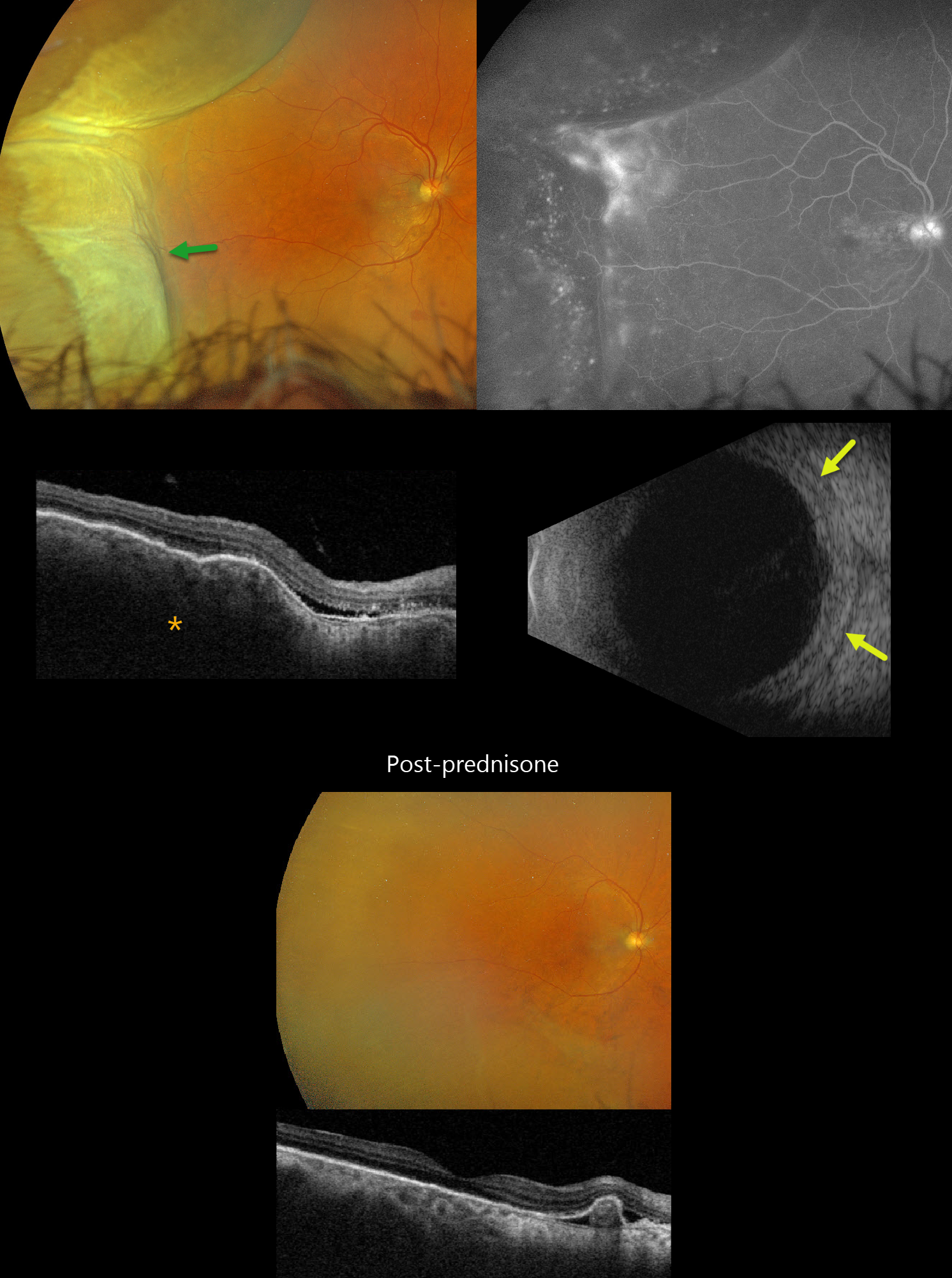
Optos color RG imaging shows solid choroidal elevation involving the temporal peripheral retina and ciliary body. Choroidal folds are evident temporally on Optos as well as on Triton swept-source OCT. The choroid is markedly thickened with trace overlying macular subretinal fluid. Fluorescein angiography shows speckled hyperfluorescence overlying the areas of peripheral choroidal elevation, more diffuse leakage along its posterior extent, late disc staining, and ill-defined submacular leakage. B-scan ultrasonography shows a diffusely thickened choroid with hyporeflective fluid posterior to the sclera (T-sign).
Oral prednisone 40mg daily was started. One week later, symptoms had improved and the temporal mass-like lesion completely resolved. OCT scanning shows choroidal fold resolution and thickening. The patient has been stable since tapering off oral prednisone, and further work-up with rheumatology found no evidence of underlying rheumatologic disease.
Learning Points:
Posterior scleritis can present either as a diffuse thickening of the entire posterior sclera or as a nodular type with localized scleral thickening (Agrawal et al, Retina 2016;36:392-401). Nodular inflammation appears as a posterior pole subretinal yellowish mass lesion, often with overlying serous fluid and chorioretinal folds. B-scan ultrasonography may show fluid in the Tenon space (“T” sign). Unlike anterior scleritis, the eyes are usually quiet and pain-free. Initial treatment is usually with oral non-steroidal anti-inflammatories or corticosteroids, although patients may also require immunosuppressive therapy. For a large case series of posterior scleritis, see McCluskey et al, Ophthalmology 1999;106:2380-2386.