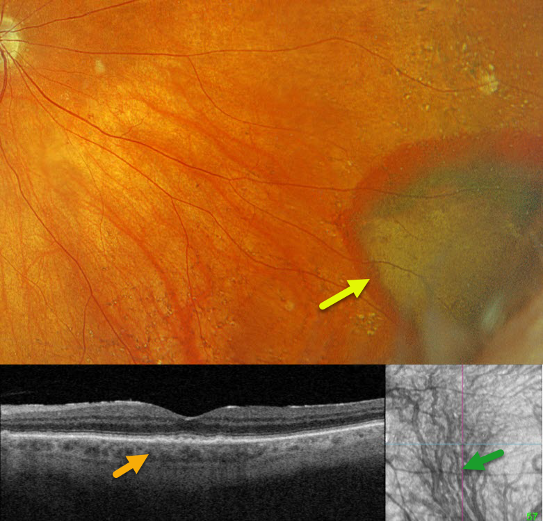
This 82YO male was referred for asymptomatic blood in his right eye. Vision was 20/40 OD.
Optos color RGB imaging shows yellow and red subretinal blood in the inferonasal midperiphery. Triton swept-source OCT shows a relatively thickened choroid for an 82YO, and choroidal en face shows an abnormal inferior dilated choroidal vortex system which extends across the horizontal meridian and drains the entire macula.
Learning Points:
Ectopic choroidal neovascularization (CNV) can often masquerade as a choroidal mass or uveal melanoma. It can also cause extensive choroidal/suprachoroidal, subretinal, and vitreous hemorrhage, more commonly in patients who are on blood thinners. The subretinal blood often spontaneously regresses without treatment, although anti-VEGF injections can be used for symptomatic or increasing exudation. We recommended observation for our patient.
Pachychoroid, meaning “thick” choroid, is secondary to large, dilated Haller vessels. Increased oncotic/mechanical pressure from these dilated vessels can cause RPE dysfunction, serous detachments, and macular neovascularization. What defines a ‘pachy’ choroid remains quite unclear (Spaide, Retina 2021;41:231-237), since pachychoroid-related findings can be present despite a ‘normal’ macular choroidal thickness as in our patient. Spaide recently described venous overload choroidopathy, a unifying concept that better explains the underlying pathogenesis (Progress Retinal Eye Research 2022;86:100973).
The pachychoroid spectrum also includes ectopic neovascularization (aka peripheral exudative hemorrhagic chorioretinopathy), with the choroid showing progressive thickening towards the temporal periphery (Shroff et al, Retina 2021;41:1518-1525).