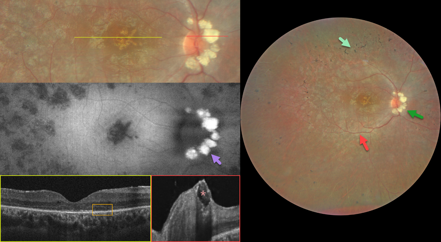
This 24YO male has a known history of retinitis pigmentosa (RP) with long-standing stable vision loss. Vision was 20/50 OD and 20/200 OS.
Color photography OD shows classic RP findings, including mid-peripheral intraretinal pigment migration (bone spicules), narrowed retinal vessels, and a pale disc. Extensive optic disc drusen (ODD) are present and markedly hyperautofluoresce. OCT shows outer retinal and RPE atrophy. A large disc druse is noted in the superficial inner portion of the disc, with a hyperreflective margin, hyporeflective core, and posterior shadowing. Similar findings were noted in his left eye (not shown).
Learning Points:
ODD are refractile calcific concretions that form anterior to the lamina cribrosa. They are thought to be extruded extracellular calcified mitochondria and may be inherited as an autosomal dominant trait. In patients with RP, they are present in about 10% of eyes on fundus photography (Grover et al, Ophthalmology 1997;104:295-298) and in 30% on EDI-OCT (Steensberg et al, J Neuroophthalmol 2024;44:483-487). ODD in isolation can cause progressive visual field defects or central vision loss, which can often confound assessment of RP-related vision loss.