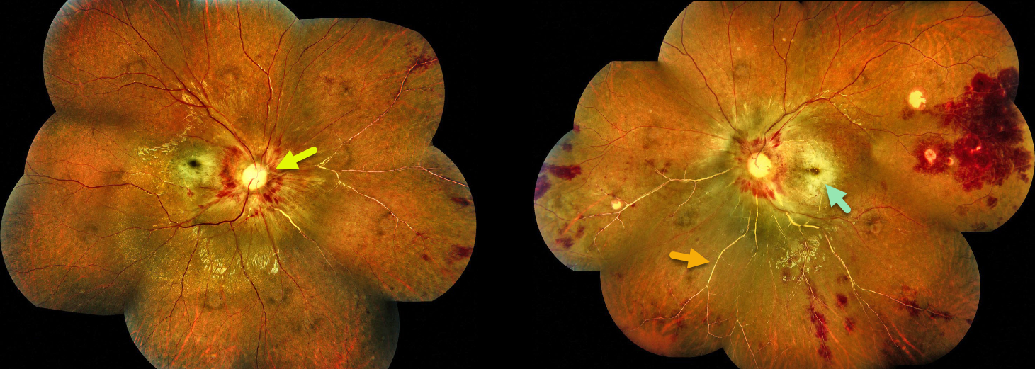
This 10YO male has a 1-year history of acute lymphoblastic leukemia with ongoing palliative treatment. He noticed decreased vision bilaterally for 3 months before presenting with light perception OU.
Fundus photography shows a bilateral cherry-red spot, pale nerves, peripapillary and peripheral retinal hemorrhages, and a white retinal arterial vascular tree.
Learning Points:
Ocular leukemic manifestations primarily result from accompanying hematologic abnormalities, including hyperviscosity, anemia, and thrombocytopenia (Soman et al, Ophthalmology Retina 2018;2:17-23). Posterior segment findings include retinal hemorrhages (sub-ILM, nerve fiber layer, or deeper, white-centered), cotton-wool spots, dilated retinal veins, and serous retinal detachment. Leukemic infiltration of the optic nerve, retina, and choroid can also occur.
Our patient’s fundus findings are most consistent with combined central retinal artery (CRAO) and central retinal vein (CRVO) occlusions. Leukemia is a well-documented though uncommon cause of CRVO (Chen et al, Retina 2026;46:796-810), and combined CRAO-CRVO is thankfully even less common (Kim et al, Graefes Arch Clin Exp Ophthalmol 2023;261:1893–1900).
We recommended bilateral panretinal photocoagulation. Unfortunately, he was immediately lost to follow-up.