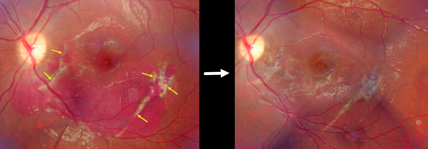
This 16YO male presented with acute vision loss in his left eye 3 days earlier following blunt trauma from a tennis ball. Vision was counting fingers.
Fundus photography shows diffuse thin subretinal blood with multiple choroidal ruptures. Observation was recommended. Vision improved to 20/200 at 6 weeks, with resolution of all blood. Mild granular pigmentary changes are present in the central macula.
Learning Points:
An acute submacular hemorrhage following blunt trauma almost always indicates an underlying choroidal rupture. As the blood clears, the rupture appears as a curvilinear or crescent-shaped yellowish-white lesion. It occurs due to rapid compression-expansion stress on Bruch’s membrane, which is less elastic and has less tensile strength than the sclera, leading to its fracture. The final vision is usually good unless the rupture extends through the macular center. These patients need long-term follow-up because of the increased risk of macular neovascularization.