This 49YO female complained of 2 months of distorted and blurred vision in her right eye and was referred for a possible uveal malignant melanoma (MM). Vision was 20/200 OD and 20/40 in her normal OS.
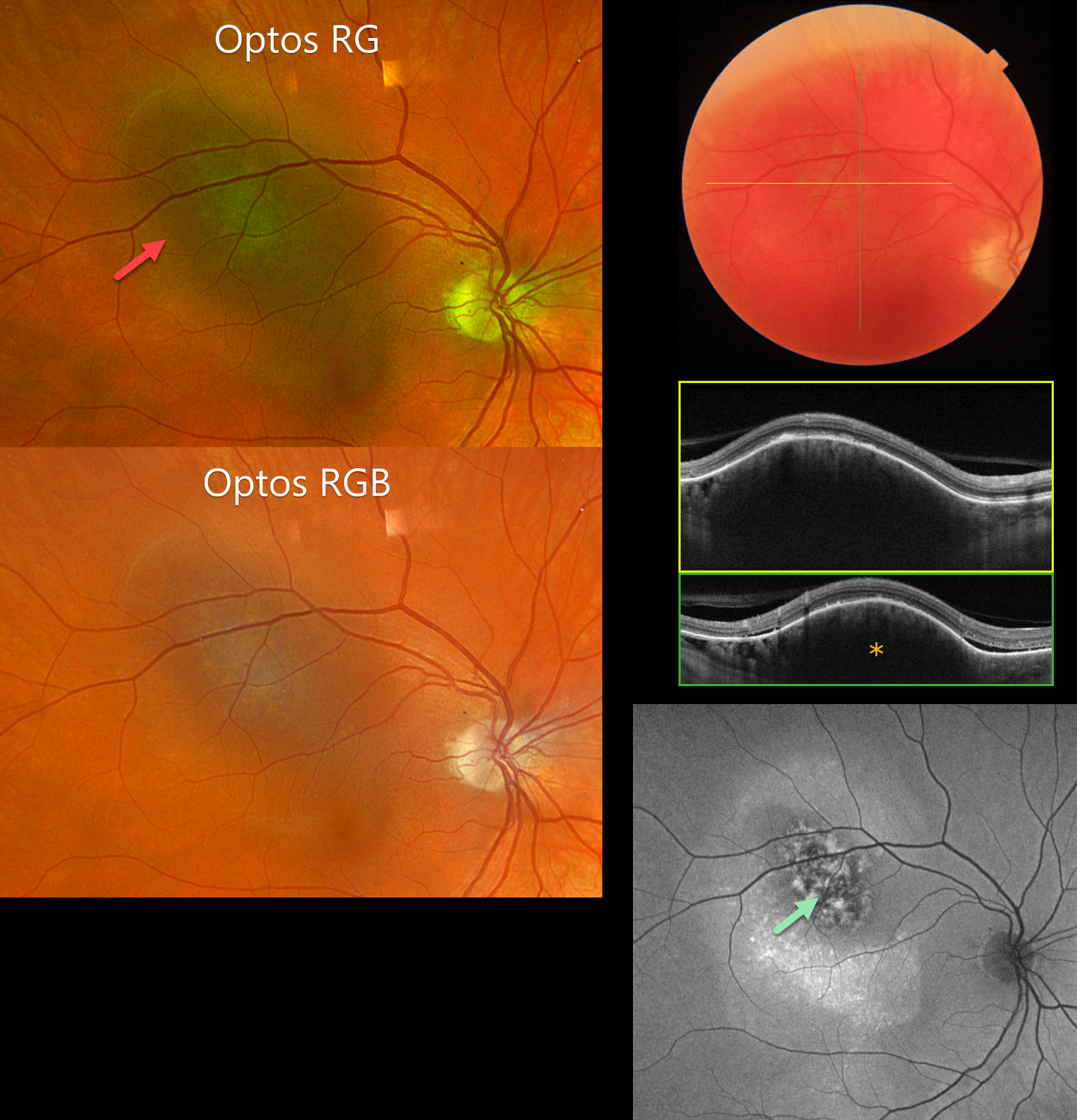
Optos color RG imaging shows a hyperpigmented subretinal lesion measuring 10.8×8.6 mm centered beneath the superotemporal arcade. However, both RGB imaging and Triton color photography reveal that this lesion is orange. A hyporeflective 1.4mm thickened choroidal lesion is noted on OCT scanning with mild subfoveal fluid. Fundus autofluorescence (FAF) shows the lesion to have a central mottled hyper- and hypo-FAF appearance, with surrounding hyper-FAF extending inferiorly.
Learning Points:
Isolated choroidal hemangiomas are benign vascular lesions. Although they have no malignant potential, they can cause vision loss from exudation leading to subretinal and intraretinal fluid, which can be treated with photodynamic therapy (see Tsipursky et al., Surv Ophthalmology 2011;56:68-85).
This lesion was initially misdiagnosed as a uveal malignant melanoma based on the hyperpigmented appearance on Optos RG imaging. This has also been reported in the literature (Becker et al, OSLIR 2023;54:292-296). These images are generated from a red (635nm) and green (532nm) laser. This creates a greenish tint, which distorts the true fundus color. In our patient, this was quite dramatic, with the true orange color of the hemangioma only evident in the Triton image. A newer Optos true color RGB unit has recently been introduced, which maintains the ability to view the separate RG channels but also generates a true-to-life color image.
Photodynamic therapy was performed for the foveal fluid. Six weeks later, the fluid completely resolved and vision remained stable at 20/100. We will continue to follow her closely.