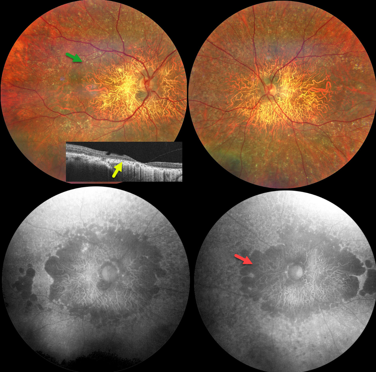
This 42YO male has a long history of night blindness, difficulty adapting to bright light, and poor vision. He had been advised elsewhere to have cataract surgery. Ocular family history was negative. Vision was counting fingers OU, and slit lamp examinations were normal.
Fundus imaging shows extensive pigmentary degeneration with subretinal crystalline deposits involving both posterior poles. OCT shows severe central and outer retinal atrophy, with more eccentric outer retinal atrophy. Fundus autofluorescence (FAF) shows diffuse hypo-FAF throughout each posterior pole with a patchier pattern of hyper- and hypo-FAF more peripherally.
Learning Points:
Bietti crystalline dystrophy is an autosomal recessive ocular disorder that affects both the anterior and posterior segments. Clinical features include crystalline deposits in the retina, cornea, and rarely the crystalline lens, as well as retinal pigment epithelial clumping and atrophy (Saatci et al, Clinical Ophthalmology 2023;17:953-967). Mutations in CYP4V2 causing dysfunctional lipid metabolism have been implicated in its pathogenesis. Although no definitive treatments exist, anti-VEGF therapy can be used for secondary macular neovascularization.