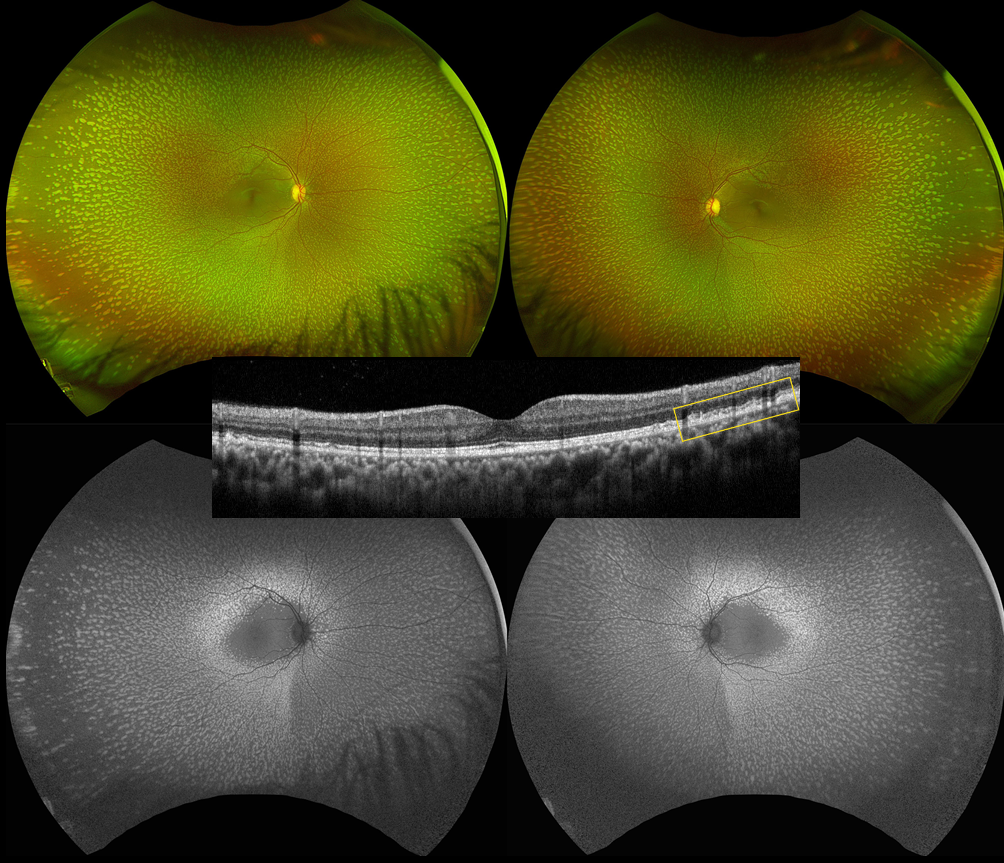
This healthy 17YO female presented for a routine ophthalmic examination. There were no ocular complaints or ocular family history. Vision was 20/20 OU.
Optos color RG imaging shows symmetrical multiple discrete yellow-white flecked lesions that spare the maculas. On OCT, the flecks appear as hyperreflective outer retinal deposits that extend into and distort the overlying ellipsoid zone. These lesions markedly hyperautofluoresce.
Learning Points:
Initially reported by Aish and Dajani (BJO 1980;64;652-659), benign familial fleck retina (BFFR) is an extremely rare autosomal recessive disorder in a heterogeneous group of flecked retina syndromes. The BFFR gene encodes for a group V phospholipase A2 (PLA2G5). Despite the dramatic fundus appearance, affected individuals have normal acuity, visual fields, and retinal function, including electrophysiology.
Other flecked retinal disorders include fundus albipunctatus, fundus flavimaculatus, familial drusen, retinitis punctata albescens, and fleck retina of Kandori. These disorders can cause night blindness, central visual problems, abnormal ERG, and abnormal perimetry. The ERG helps distinguish BFFR from other flecked retina syndromes such as retinitis punctata albescens and fundus albipunctatus, as scotopic ERG responses are decreased in the latter conditions but normal in BFFR.