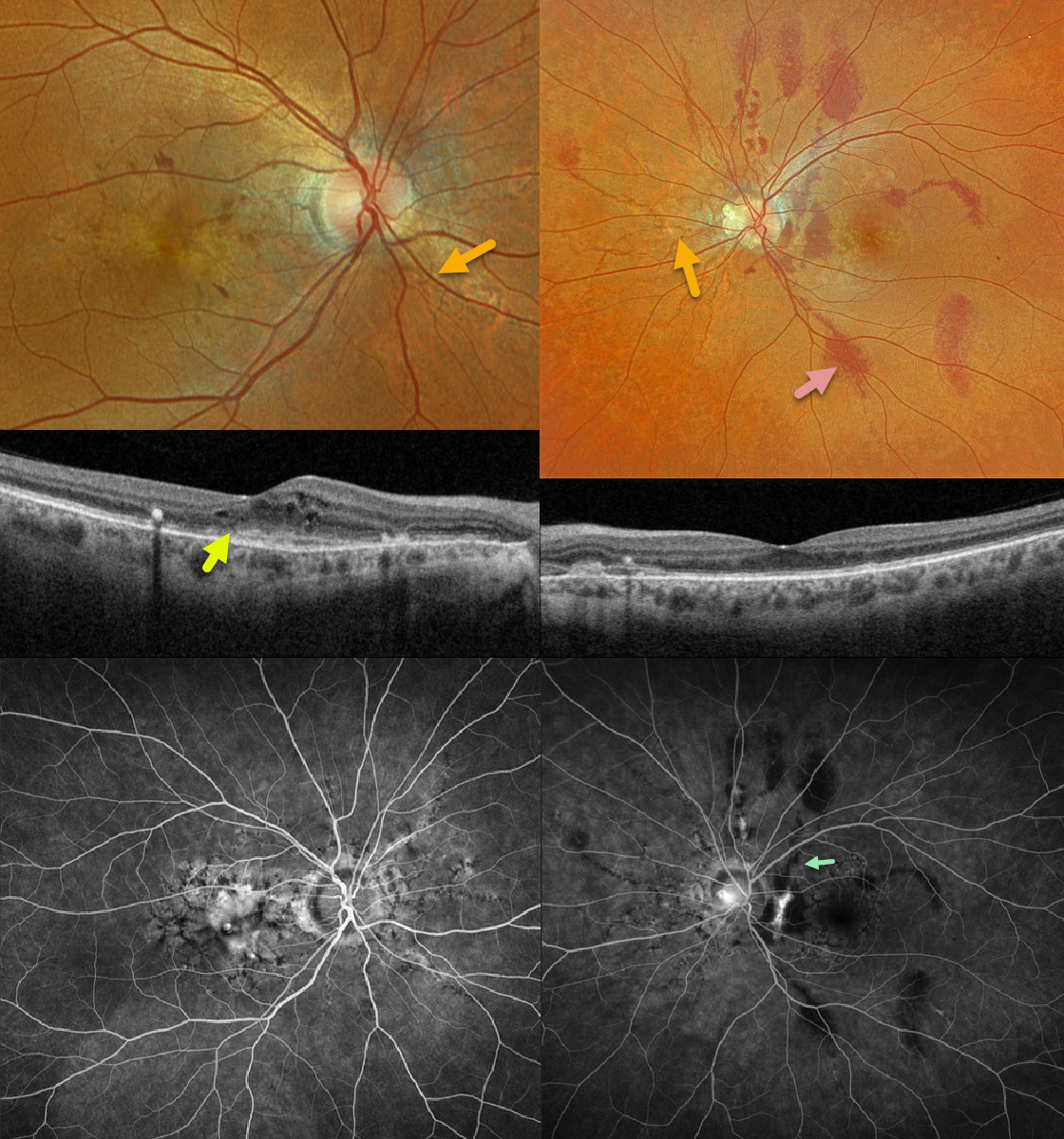
This 45YO female has a history of osteogenesis imperfecta (OI) and complained of several weeks of central vision loss in her right eye. Two months earlier, she was hospitalized following a fall in which she hit the left side of her face, resulting in a concussion and intracranial hemorrhage. Vision was counting fingers OD and 20/20 OS.
Optos color RGB imaging shows bilateral angioid streaks with a peau d’orange appearance to the posterior poles. There are numerous small deep vs subretinal hemorrhages scattered throughout the right macula. Triton swept-source OCT shows cystic retinal thickening. Multiple atypical subretinal hemorrhages mostly parallel the radiating angioid streaks in her left eye. Fundus fluorescein angiography shows diffuse submacular staining OD, a speckled pattern of hypofluorescence from the angioid streaks OU, and blockage from the subretinal blood OS.
Learning Points:
Doyne first described angioid streaks in 1889 as irregular, orange-yellow, crack-like dehiscences in Bruch’s membrane, associated with atrophic degeneration of the overlying RPE. The term “angioid” derives from its resemblance to blood vessels. Although they have historically been mistakenly associated with numerous conditions, the only truly clinically relavent association is with pseudoxanthoma elasticum (PXE; Nadelmann et al, Eye 2023;37:1596-1601). Patients are at significant risk for vision loss from MNV or pattern dystrophy-like changes (Murro et al, Graefe’s 2020;258:1881-1892).
Even minor trauma with angioid streaks can cause subretinal hemorrhaging and choroidal ruptures (Agrawal et al, JAMA Ophthalmology 2017;135(3):e165466). Unlike typical choroidal ruptures, which are usually curvilinear with the nerve, lesions from brittle Bruch’s membrane in PXE appear as numerous irregular ruptures, usually radiating from the nerve. The subretinal hemorrhages in our patient’s left eye were felt to be avascular, unlike the macular neovascularization (MNV) in her right eye.
OI is a genetic disorder that primarily affects type I collagen, leading to bone fragility and various extraskeletal manifestations, including vascular fragility. Ocular manifestations include blue sclerae, increased risk of retinal detachment, and scleral thinning. There is no published association between OI and angioid streaks, other than a single case report of classic PXE-like findings in a patient with OI (Banerjee et al, BMJ Case Rep 2023;16:3256366.doi:10.1136/bcf-2023-256366). However, genetic testing was not reported, so we cannot determine whether this was a chance association with PXE or isolated to OI. Our patient did not have genetic testing due to cost. We suspect our patient has PXE.
Monthly intravitreal injections were started for the MNV in her right eye. Vision improved to 20/30 with complete resolution of all exudation and blood.