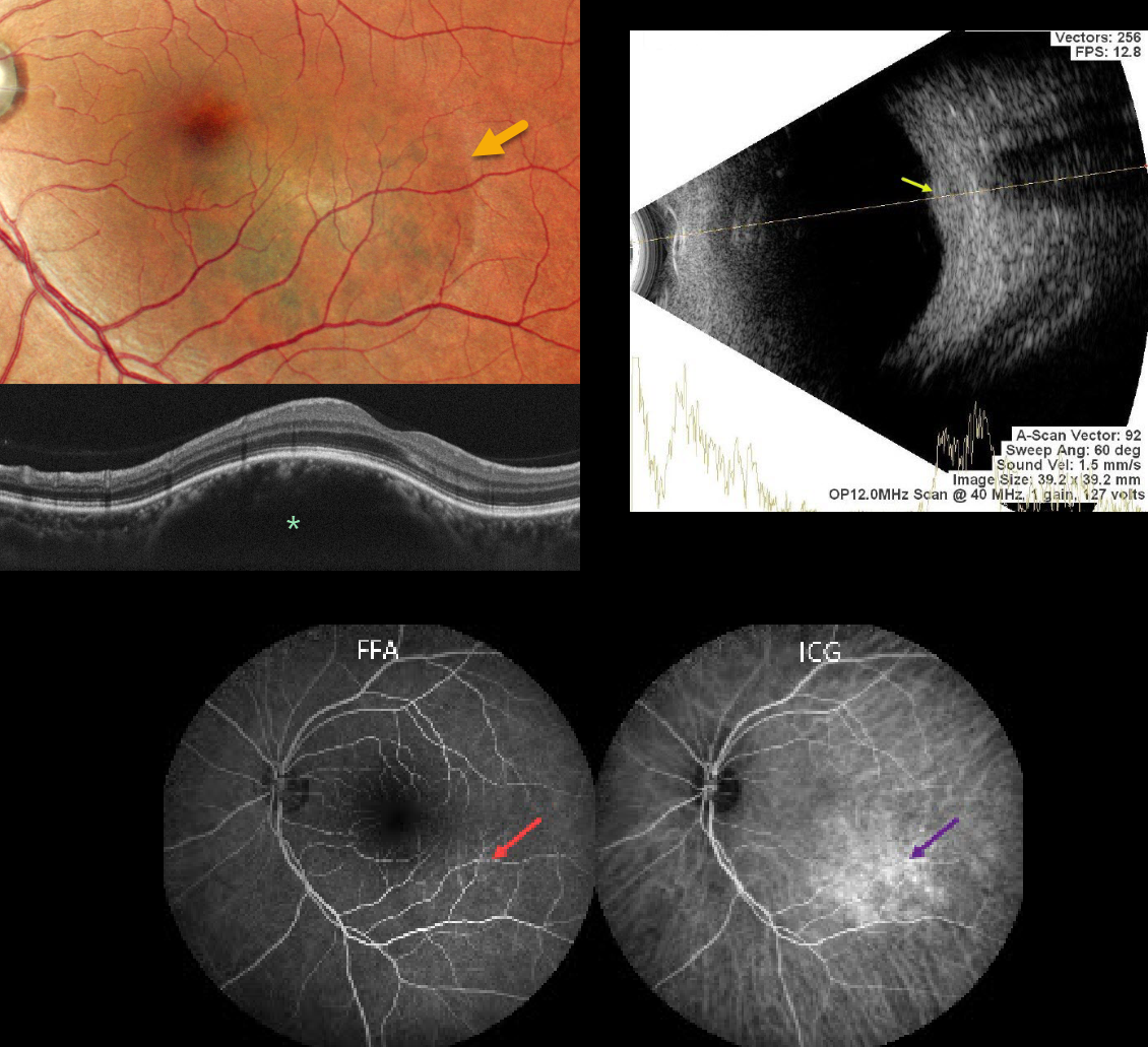
This 48YO female presented with 6 months of mild blurred vision in her right eye. She was diagnosed elsewhere with a choroidal granuloma and referred to us for further evaluation. Vision was 20/20 OD and 20/25 in her normal OS.
Pseudocolor SLO imaging shows a well-demarcated, round, dome-shaped orange-red choroidal lesion centered in the infotemporal macula. OCT shows a smooth, dome-shaped, elevated choroidal lesion with a gently sloping anterior contour. The overlying retina appears normal. The lesion shows early autofluorescence with late leakage on fluorescein angiography and is somewhat better imaged with indocyanine green angiography (ICGA), although late-phase ICGA showing tumor washout was not possible due to patient vomiting and allergic hives. B-scan ultrasonography shows a smoothly thickened choroidal lesion with high internal reflectivity and homogeneous internal acoustic characteristics.
We recommended observation due to the lack of symptoms or exudation.
Learning Points:
Choroidal hemangioma is an uncommon benign hamartomatous disorder that presents as either a circumscribed, orange-red, round choroidal lesion (as in our patient), which is almost always isolated and nonsyndromic, or a diffuse form, which is usually part of Sturge-Weber syndrome. Although there is no malignant potential, it can cause vision loss from exudation leading to subretinal and intraretinal fluid, which is best treated with photodynamic therapy (see Yang et al., Surv Ophthalmology 2025;70:389-400).