This previously healthy 34YO female presented with recent bilateral vision loss. She recalled having blood pressure issues during her last pregnancy. Vision was hand motion OD and 20/80 OS.
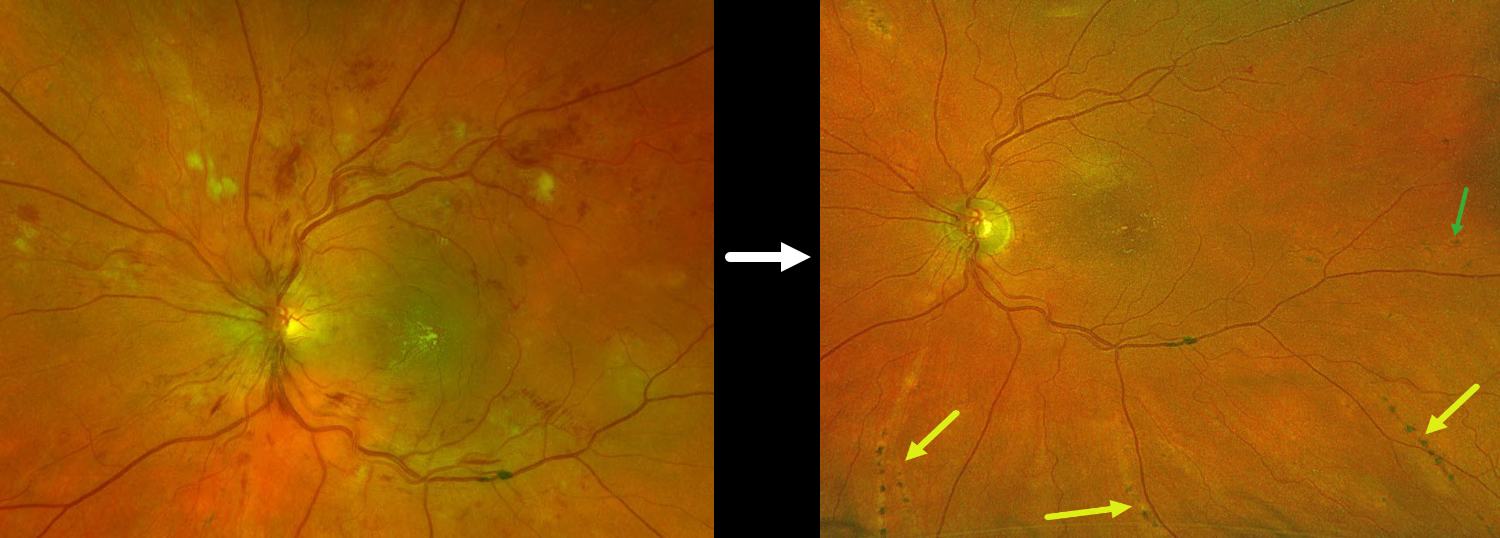
Optos color RG imaging of her left eye shows disc swelling, scattered inner retinal hemorrhages, cotton wool spots (CWS), and a nasal macular lipid star. Similar findings were noted in her right eye (not shown). Blood pressure was 292/208, and she was immediately referred to the emergency room for malignant hypertension.
She was then lost to follow-up when she presented 4 years later. Vision was 20/20 bilaterally. Optos color RG imaging shows radiating linear areas of pigment loss and clumping, with focal subretinal pigment clumps.
Learning Points:
Malignant hypertension, defined as blood pressure above 180 systolic and/or 120 diastolic, is a life-threatening medical emergency. Eye doctors are often uniquely positioned to diagnose this condition (Shukla et al, Indian J Ophthalmol 2025;73:1552-1555). Patients will often present with bilateral optic nerve swelling, flame-shaped retinal hemorrhages, CWS, and, with more chronic disease, lipid precipitates in the nasal macular outer plexiform layer.
Acute hypertensive choroidopathy outside the setting of pre-eclampsia, in our experience, is quite rare. Independent of the disc and neurosensory retinal findings, patients present with localized multifocal serous retinal detachments (de Venecia and Jampol, Arch Ophthalmol 1984;102:68-73). With blood pressure control, these often resolve with minimal funduscopic changes. Focal (Elschnig spots) or linear (Siegrist streaks) choroidal pigmentary changes, as seen in our patient, may indicate more severe disease.