Is your case great enough to be selected for our Wall of Fame? If so, we’ll post your name and case here for the world to see!
4/25: Anand Temkar and Manish Nagpal
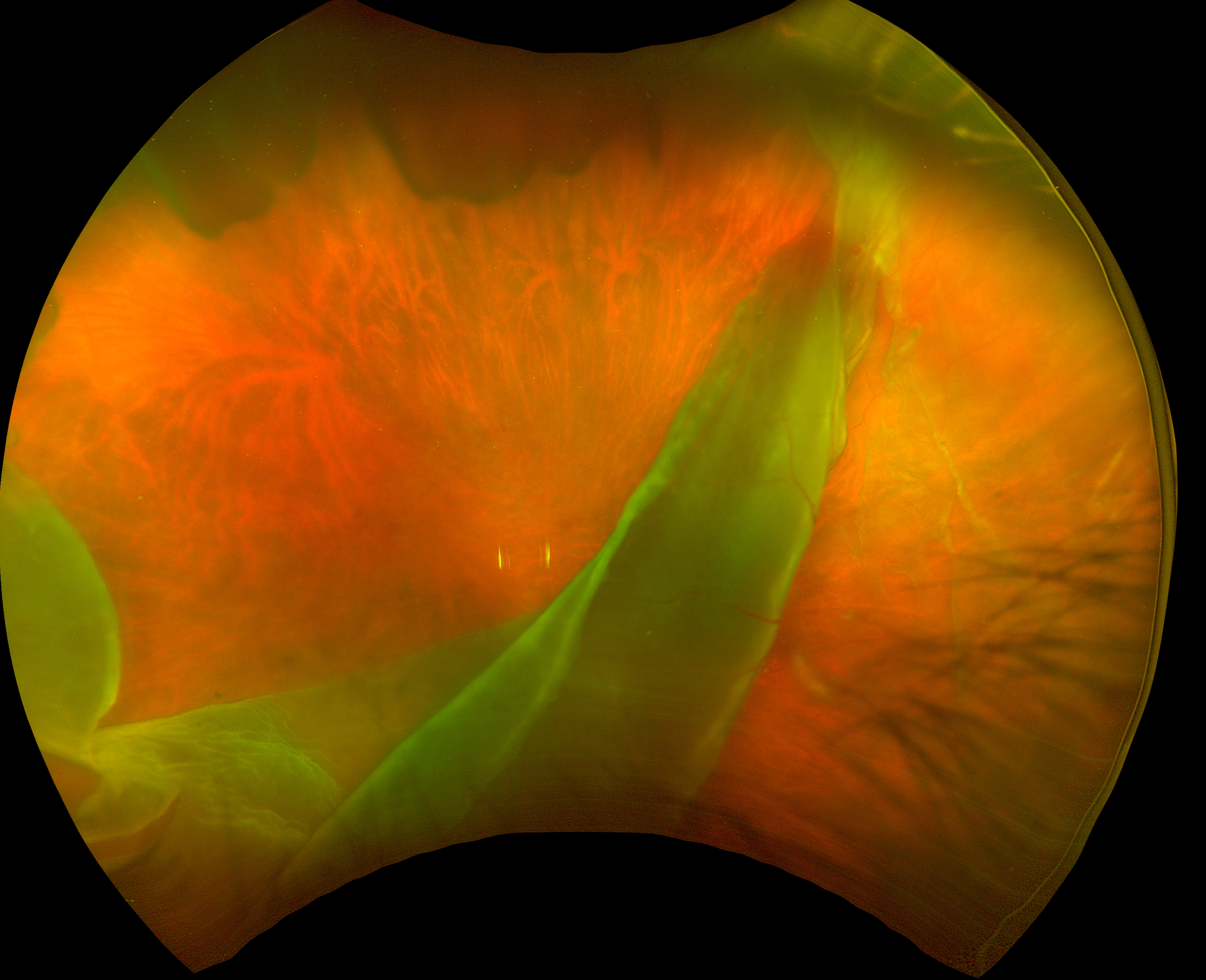
Tamoxifen retinopathy
3/25: Anand Temkar and Manish Nagpal
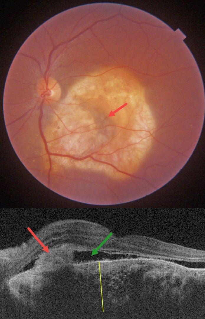
Total retinal detachment from giant macular hole
2/25: Vaibhav Sethi
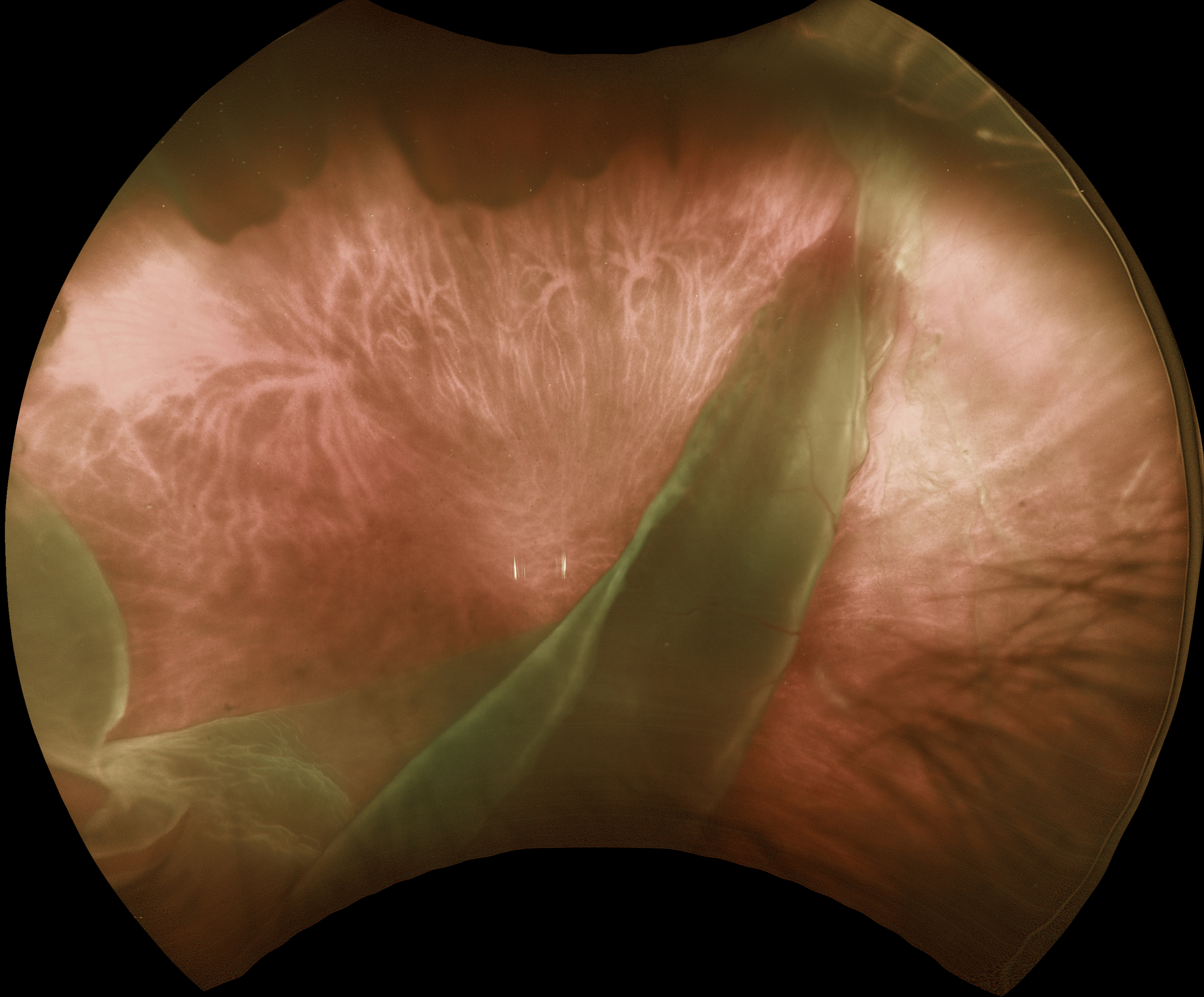
Degenerative retinoschisis
1/25: Anjana Mirajkar and Manish Nagpal
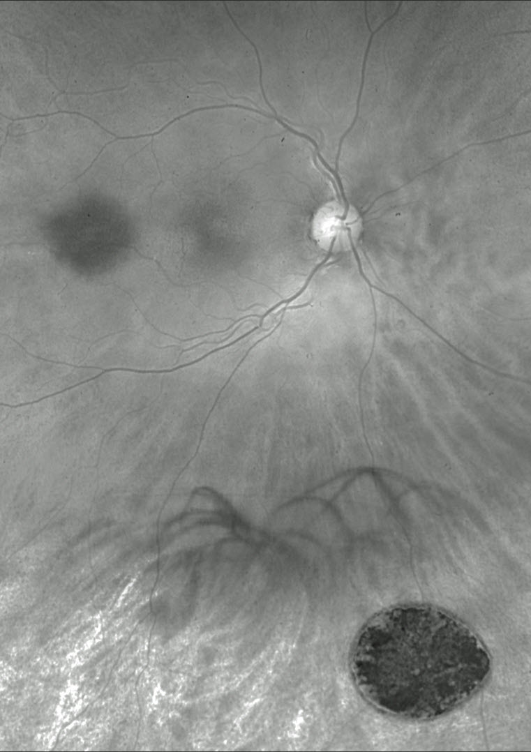
Giant tear retinal detachment
12/24: Sharat Hegde
Diffuse unilateral subacute neuroretinitis (DUSN)
11/24: Anand Temkar and Manish Nagpal
Unilateral anemic retinopathy with optic nerve edema
10/24: Evgenii Chernov
Best disease
9/24: Anjana Mirajkar and Manish Nagpal
Intraocular foreign body
8/24: Anjana Mirajkar and Manish Nagpal
Pseudopapilledema from leukemic optic nerve infiltration
7/24: Justin Grassmeyer, Ambar Faridi, and Brittany Heckerman
Lattice degeneration
6/24: Ogugua Okonkwo, Adekunle Olubola Hassan, Ayodele Harriman, Ogochukwu Sibeudu, and Idris Akintayo Oyekunle
Tuberculous serpiginous-like choroiditis
5/24: Seif Anwar
Inherited retinal disease
Sengül Özdek and Ece Özdemir Zeydanlı
Case of the Year winner!
Retinal Arteriovenous malformation
4/24: Kanwaljeet Harjot Madan
Shaken baby syndrome
3/24: Ayushi Gupta and Vishal Agrawal
Benign familial fleck retina
2/24: Will Gibson
Retinal venous malformation
12/23: Anjana Mirajkar and Manish Nagpal
Central retinal artery occlusion with cilioretinal sparing
11/23: Anjana Mirajkar and Manish Nagpal
Harada’s disease
10/23: Will Gibson
Posterior scleritis with chorioretinal folds
9/23: Shivraj Tagare and Nishant Maindargi
Valsalva retinopathy drained with YAG laser
8/23: Mohammad Abbas
Laser-induced retinal break and vitreous hemorrhage
7/23: Joe Yuenpang Cheung
Group-type congenital pigmented nevi of the RPE (bear tracks)
6/23: Gil Calvão-Santos and Keissy Sousa
Waardenburg Syndrome
5/23: Natasa Draca and Ratko Lazic
Tattoo-induced Uveitis
4/23: Veronika Matello and Barbara Parolini
Case of the Year winner!
The EmPuzzled Eye
3/23: Emma Oreskovic and Natasha Draca
Optic nerve pit
2/23: Anjana Mirajkar
Giant RPE tear
11/22: Veronika Matello and Barbara Parolini
Choroidal hemangioma widefield OCT
8/22: Nivesh Gupta
Ischemic central retinal vein occlusion
7/22: Omar Mulki:
Choroidal osteoma
3/22: Erdem Dinç
Is temporary ILM flap sufficient for MH closure?
1/22: Veronika Matello and Barbara Parolini
Case of the Year winner!
Choroidal transplant for subfoveal AMD MNV
12/21: Otis Hertsenberg
Optic nerve coloboma with pit
11/21: David Kilpatrick
Frosted branch angiitis from sarcoidosis
10/21: Kumar Chugani
Coats disease


